. 2005 Dec;89(6):4364-73.
doi: 10.1529/biophysj.105.064642. Epub 2005 Sep 8.

Molecular identification and reconstitution of depolarization-induced exocytosis monitored by membrane capacitance

Affiliations

Molecular identification and reconstitution of depolarization-induced exocytosis monitored by membrane capacitance

Roy Cohen et al. Biophys J. 2005 Dec.

Abstract

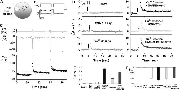
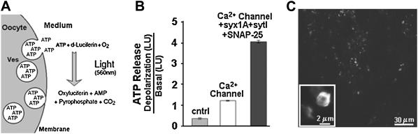
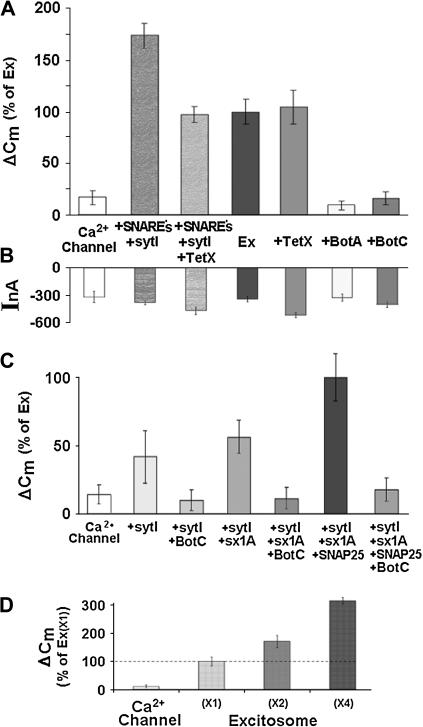
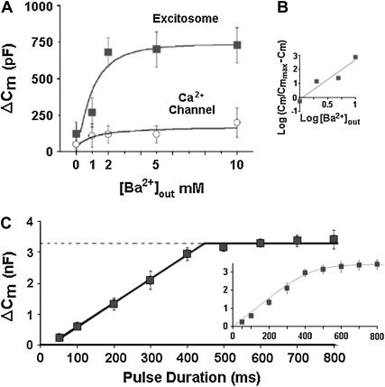
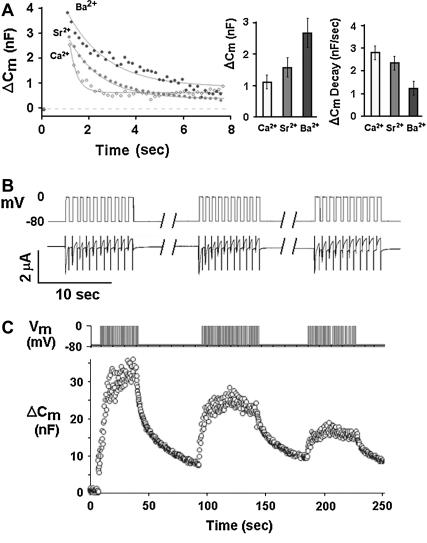
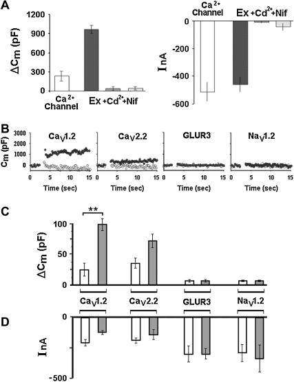
Regulated exocytosis of neurotransmitters at synapses is fast and tightly regulated. It is unclear which proteins constitute the "minimal molecular machinery" for this process. Here, we show that a novel technique of capacitance monitoring combined with heterologous protein expression can be used to reconstitute exocytosis that is fast (<0.5 s) and triggered directly by membrane depolarization in Xenopus oocytes. Testing synaptic proteins, voltage-gated Ca2+ channels, and using botulinum and tetanus neurotoxins established that the expression of a Ca2+ channel together with syntaxin 1A, SNAP-25, and synaptotagmin was sufficient and necessary for the reconstitution of depolarization-induced exocytosis. Similar to synaptic exocytosis, the reconstituted release was sensitive to neurotoxins, modulated by divalent cations (Ca2+, Ba2+, and Sr2+) or channel (Lc-, N-type), and depended nonlinearly on divalent cation concentration. Because of its improved speed, native trigger, and great experimental versatility, this reconstitution assay provides a novel, promising tool to study synaptic exocytosis.


Figures

FIGURE 1
FIGURE 1
Reconstitution of depolarization-induced exocytosis in oocytes by expression of exogenous proteins. (A) Diameter and capacitance of plasma membrane and cortical granules in Xenopus oocytes. (B) Effect of depolarization on membrane capacitance in a Xenopus oocyte expressing a voltage-gated Ca2+ channel. (Upper trace) Depolarizing voltage command (see Methods), consisting of depolarization from a holding potential of −80 mV to 0 mV for 2 × 500 ms, separated by 100 ms at −80 mV). (C) Continuous monitoring of membrane capacitance showing the effect of depolarization on membrane capacitance (Cm) in an oocyte expressing, Ca2+ channel, Lc-type Ca2+ channel subunits α11.2, β2A, α2δ; SNAREs: syntaxin 1A, SNAP-25, and synaptotagmin; representative original traces of voltage (upper) current (middle) and Cm (lower). The vertical dashed line indicates the time at which Cm was read for comparison with the baseline Cm before depolarization. (D) Monitoring Cm in oocytes expressing different proteins. Control, uninjected oocytes; other panels, oocytes expressing heterologously synaptic proteins: synaptic proteins, syntaxin 1A, SNAP-25, synaptobrevin, and synaptotagmin, Ca2+ channel, Lc-type Ca2+ channel subunits α11.2, β2A, α2δ; SNAREs: syntaxin 1A, SNAP-25 and synaptobrevin; synaptotagmin I, sytI; Ca2+ channel plus syntaxin 1A, SNAP-25 and syt I. (E) Summary: effect of depolarization on Cm. Groups as in D. ΔCm, depolarization-induced change of membrane capacitance; bars show mean ± SD (n = 13). (F) Effect of depolarization on membrane current. Depolarization-induced membrane current (InA; mean ± SD, n = 13 each). Data show that the Cm changes (C) cannot be explained by membrane current, but rather reflect bona fide exocytosis.
FIGURE 2
FIGURE 2
Effect of heterologous proteins on depolarization-induced ATP release. (A) A schematic presentation of ATP luminescence assay. (B) Oocytes were depolarized by incubation in high K+-solution, and release of ATP was determined via a luminescence assay (see Methods). ATP release paralleled the Cm changes (cf. Fig. 1, A and B), confirming that Cm changes indeed reflect fusion of cytoplasmic vesicles with the plasma membrane. (C) Heterologously expressed synaptotagmin is localized in cortical granules. Confocal image of oocytes injected with cRNA for a GFP-synaptotagmin 1-fusion protein.
FIGURE 3
FIGURE 3
Dissecting out the role of synaptic proteins in reconstituted depolarization-induced exocytosis. (A) Effects of botulinum toxins (BotC and BotA) and tetanus toxin (TetX) on depolarization-induced capacitance changes. Lc-type Ca2+ channel (Cav1.2); SNAREs (syntaxin 1A, synaptobrevin, and SNAP-25) and synaptotagmin I; Ex, Ca2+ channel + syntaxin 1A + SNAP-25, were expressed at a level corresponding to “×1” in Fig. 3 D. Data show that SNAP-25 and syntaxin 1A are absolutely required for depolarization-induced exocytosis. In contrast, synaptobrevin is not required, although it increases exocytosis efficiency. (B) Effects of botulinum toxins and tetanus toxin on depolarization-induced currents. Current amplitudes and Cm changes (A) are not correlated. (C) Stepwise molecular reconstitution of depolarization-induced exocytosis. Synaptic proteins as above, and Lc-type Ca2+ channel (Cav1.2). Data show that syntaxin 1A and SNAP-25 are absolutely required for depolarization-induced exocytosis. The changes in Cm in the presence of the toxins are similar to the step Cm expressed by the channel alone. Data suggest that the synaptic proteins comprised by the excitosome are necessary and sufficient to specifically reconstitute depolarization-induced exocytosis against a very small background in Xenopus oocytes. (D) Quantitative correlation between reconstituted depolarization-induced exocytosis and expression of excitosome proteins. Coexpression of graded amounts of the synaptic proteins syntaxin 1A, synaptotagmin I, and SNAP-25 together with a fixed amount of Lc-type voltage-gated Ca2+-channel subunits that comprise the excitosome (see Methods).
FIGURE 4
FIGURE 4
Quantitative characterization of reconstituted depolarization-induced exocytosis. (A) Dependence of depolarization-induced ΔCm on extracellular Ba2+ concentration. Depolarization from −80 mV to 0 mV for 2 × 500 ms 100 ms apart in oocytes expressing Lc-type channel without (lower trace) or with (upper trace) the synaptic proteins syntaxin 1A, SNAP-25, and synaptotagmin I, as in Fig. 1. (B) Hill plot. Data (from A) show saturation of the Cm effect >2 mM, and a nonlinear concentration-dependence of ΔCm with an estimated Hill coefficient of nH ≈ 2.8. (C) Dependence of depolarization-induced ΔCm on depolarization duration. Depolarization from −80 mV to 0 mV for indicated times in 5 mM Ba2+. A linear correlation observed with pulse duration for longer depolarization periods, saturation at ∼400 ms and half-maximal capacitance changes are reached at a depolarization time of ∼250 ms. A straight line fitted to the data between 50 and 400 ms had a slope of 7.7 pF ms−1 (R2 = 0.998) corresponding to ∼600,000 vesicles at saturation. The results could be fitted less well with a single exponent (inset) showing a time constant of 101.05 ± 12 ms corresponding to an initial rate of 6 × 10⁶ vesicles/s (inset). The saturation in the capacitance jumps defines a readily releasable pool of ∼106–107 vesicles (see 31).
FIGURE 5
FIGURE 5
Differential effects of Ca2+, Sr2+, or Ba2+ on depolarization-induced Cm changes, and vesicle depletion by repetitive stimulation (A) Differential effects of Ca2+, Sr2+, or Ba2+ on depolarization-induced exocytosis. Oocytes expressing Cav1.2, syntaxin 1A, syt-1, and SNAP-25 (excitosome). (Left panel) Original traces showing time course of Cm upon depolarization, fitted by simple exponentials; (middle panel) instantaneous depolarization-induced Cm increase in Ca2+, Sr2+, and Ba2+ (mean ± SE, n = 10, each); and (right panel) rate of compensatory Cm decrease (mean ± SE, n = 10, each). As compared to Ca2+, the instantaneous Cm increase was greater in Ba2+ (middle panel), and the compensatory Cm decrease was slower (right panel). In Sr2+, the respective values were between those of Ca2+ and Ba2+. (B) Current amplitude during repeated trains of membrane depolarization. Oocytes were depolarized by three trains composed of 10 depolarizations from −80 to 0 mV 1 s each (upper panel). Depolarization-induced inward current inactivated during trains whereas no run-down of current amplitude was observed from one train to the other (lower panel). (C) Exhaustibility of depolarization-induced capacitance changes. Repeated depolarization pulses from −80 mV to 0 mV for 1 s each (upper trace) and the associated Cm changes (lower trace). Data are consistent with depletion and only partial replenishing of a pool of releasable vesicles.
FIGURE 6
FIGURE 6
Dissecting out the role of the Ca2+ channel in reconstituted depolarization-induced exocytosis (A) Effect of Ca2+ channel inhibitors on reconstituted depolarization-induced exocytosis. Oocytes were expressing either the Lc-type Ca2+ channel alone (leftmost bar), or together with the synaptic proteins syntaxin 1A (sx1A), synaptotagmin I (syt I), and SNAP-25 (other three bars). Depolarization-induced changes of capacitance (ΔCm) and current amplitude (I) in 5 mM Ba2+. The effects of the Ca2+ channel blockers Cd2+ (200 μM) and nifedipine (10 μM) show that depolarization-induced exocytosis absolutely requires the presence and functionality of a voltage-gated Ca2+ channel. (B) Effect of channel type on depolarization-induced Cm changes. Time course of Cm in oocytes expressing various voltage-gated channels (L-, N-type Ca2+ channels, GluR3 receptor, or brain-type II Na+ channel; see Methods) without (open circles) or with SNAP-25, syntaxin 1A, and synaptotagmin 1 (solid circles); bath containing 5 mM Ba2+ (with Ca2+ channels and GluR3) or 50 mM Na+ (with Na+ channel). Depolarization from −80 mV to 0 mV for 2 × 500 ms. (C) Effect of channel type on size of depolarization-induced Cm increase. Same protocol as in (A) (open bars) channel only; (solid bars) channel plus SNAP-25, syntaxin 1A, and synaptotagmin 1. Mean ± SD from 10 oocytes, each. Cav1.2 versus Cav1.2 + synaptic proteins (**), P < 0.0015; Cav2.2 versus Cav2.2 + synaptic proteins P* < 0.1 in Student's t-test. (D) Effect of channel type on depolarization-induced current changes. Same protocol as in A and B.

Similar articles

Cited by

References

    1. Katz, B., and R. Miledi. 1969. Spontaneous and evoked activity of motor nerve endings in calcium ringer. J. Physiol. 203:689–706. - PMC - PubMed
    1. Mayer, A. 2002. Membrane fusion in eukaryotic cells. Annu. Rev. Cell Dev. Biol. 18:289–314. - PubMed
    1. Jahn, R., T. Lang, and T. C. Sudhof. 2003. Membrane fusion. Cell. 112:519–533. - PubMed
    1. Martin, T. F. 2003. Tuning exocytosis for speed: fast and slow modes. Biochim. Biophys. Acta. 1641:157–165. - PubMed
    1. Weber, T., B. V. Zemelman, J. A. McNew, B. Westermann, M. Gmachl, F. Parlati, T. H. Sollner, and J. E. Rothman. 1998. SNAREpins: minimal machinery for membrane fusion. Cell. 92:759–772. - PubMed

LinkOut - more resources