. 2005 Sep;71(9):5066-76.
doi: 10.1128/AEM.71.9.5066-5076.2005.

Gene cluster responsible for validamycin biosynthesis in Streptomyces hygroscopicus subsp. jinggangensis 5008

Affiliations

Gene cluster responsible for validamycin biosynthesis in Streptomyces hygroscopicus subsp. jinggangensis 5008

Yi Yu et al. Appl Environ Microbiol. 2005 Sep.

Abstract

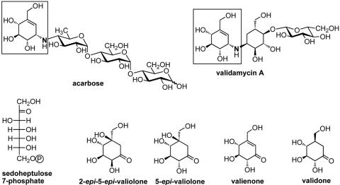
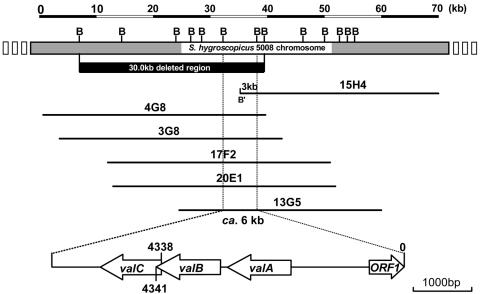
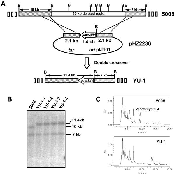
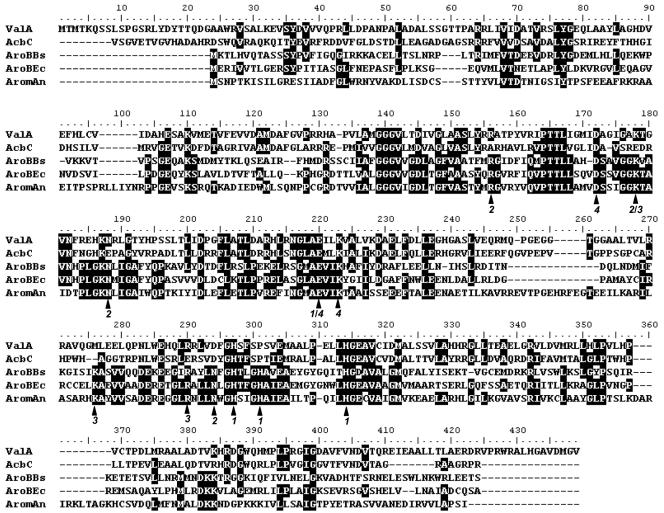
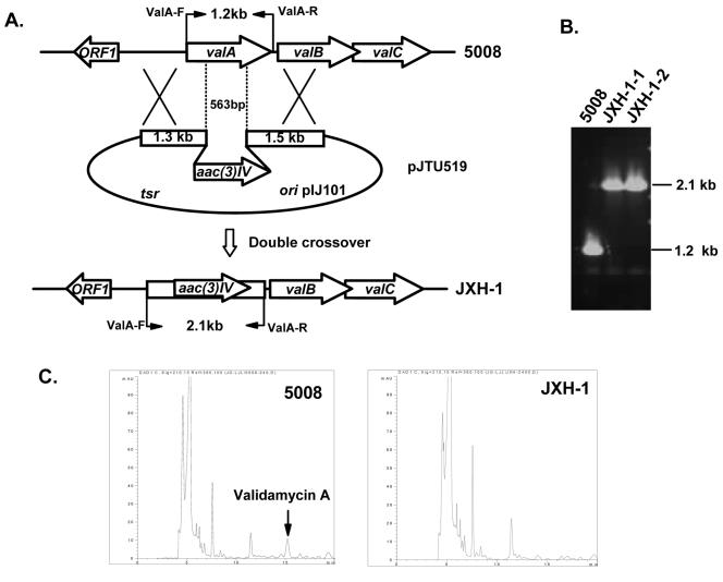
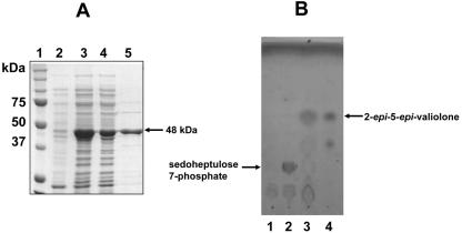
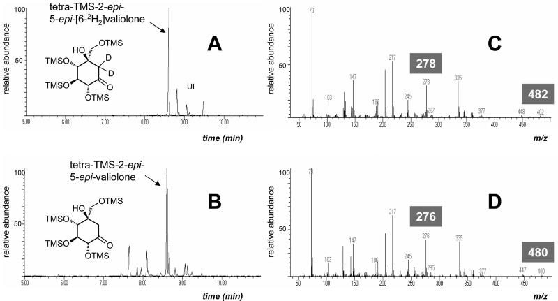
A gene cluster responsible for the biosynthesis of validamycin, an aminocyclitol antibiotic widely used as a control agent for sheath blight disease of rice plants, was identified from Streptomyces hygroscopicus subsp. jinggangensis 5008 using heterologous probe acbC, a gene involved in the cyclization of D-sedoheptulose 7-phosphate to 2-epi-5-epi-valiolone of the acarbose biosynthetic gene cluster originated from Actinoplanes sp. strain SE50/110. Deletion of a 30-kb DNA fragment from this cluster in the chromosome resulted in loss of validamycin production, confirming a direct involvement of the gene cluster in the biosynthesis of this important plant protectant. A sequenced 6-kb fragment contained valA (an acbC homologue encoding a putative cyclase) as well as two additional complete open reading frames (valB and valC, encoding a putative adenyltransferase and a kinase, respectively), which are organized as an operon. The function of ValA was genetically demonstrated to be essential for validamycin production and biochemically shown to be responsible specifically for the cyclization of D-sedoheptulose 7-phosphate to 2-epi-5-epi-valiolone in vitro using the ValA protein heterologously overexpressed in E. coli. The information obtained should pave the way for further detailed analysis of the complete biosynthetic pathway, which would lead to a complete understanding of validamycin biosynthesis.


Figures

FIG. 1.
FIG. 1.
Chemical structures of acarbose and validamycin A (top), and the initial intermediates proposed to be involved in validamycin A biosynthesis (bottom) (7). Boxed regions show the valienamine moiety shared by both compounds.
FIG. 2.
FIG. 2.
Overlapping cosmids covering genes for validamycin biosynthesis and gene organization of the 6-kb sequenced region. Top: BamHI restriction map of the ca. 70-kb contig for validamycin biosynthesis. The solid bar indicates the 30-kb deleted region in Fig. 3, which abolished validamycin production. Bottom: 6-kb sequenced region including the acbC homologue (valA). B: BamHI site. B′: regenerated BamHI site from BamHI (vector) and MboI (insert) sites.
FIG. 3.
FIG. 3.
Replacement of a 30-kb region containing the acbC homologue by aac(3)IV in strain 5008. (A) Schematic representation of the replacement of the 30 kb of DNA mediated by 2.1-kb genomic fragments flanking both sides of the 1.4-kb aac(3)IV apramycin resistance determinant in pHZ2236. The mutants generated by double crossover (YU-1-1 to YU-1-4) had a fusion of the 10-kb leftward BamHI fragment with the BglII end of the 1.4-kb aac(3)IV to form an 11.4-kb new BamHI fragment, but keep the rightward 7-kb fragment unchanged. (B) Southern hybridization using the α-[32P]dCTP-labeled 5.6 (2.1 + 1.4 + 2.1)-kb insert from pHZ2236 as the probe after genomic DNAs of the wild-type 5008 and its derivatives (YU-1-1 to YU-1-4) were digested with BamHI. (C) HPLC analysis demonstrating that validamycin A is produced by wild-type strain 5008, but not by YU-1.
FIG. 4.
FIG. 4.
Alignment of ValA with AcbC and three AroB proteins. Deduced amino acid sequences are from the following organisms: AcbC, Actinoplanes sp. (Y18523.3); AroBBs, Bacillus subtilis (M80245); AroBEc, E. coli (X03867); and AromAn, Emericella (formerly Aspergillus) nidulans (395-amino-acid DHQS domain at the N terminus of the pentafunctional AROM protein; X05204). A functional attribution of the amino acid residues indicated with black arrows, based on the analysis of the three-dimensional structure of the dehydroquinate synthase (DHQS) domain of E. nidulans (4), is given below the alignment by the following code: 1 = Co2+ binding (Zn2+ in the fungal protein instead); 2 = heptulose phosphate group binding; 3 = C-1 hydroxyl fixation; and 4 = C-4 hydroxyl fixation.
FIG. 5.
FIG. 5.
Inactivation of valA of the validamycin biosynthetic gene cluster. (A) Schematic representation of the replacement of a 563-bp internal fragment of valA with the 1.4-kb aac(3)IV. In shuttle plasmid pJTU519, aac(3)IV was inserted between 1.3-kb and 1.5-kb genomic fragments originally flanking the deleted 563-bp region. While wild-type 5008 should give a 1.2-kb PCR-amplified product, mutant JXH-1 should yield a 2.1-kb product using a pair of primers (ValA-F and ValA-R) designed for amplification of full-length valA. (B) PCR analysis of wild-type 5008 and mutant JXH-1. (C) HPLC comparison between 5008 and JXH-1. The peak corresponding to validamycin A is absent from mutant JXH-1.
FIG. 6.
FIG. 6.
(A) Sodium dodecyl sulfate-polyacrylamide gel electrophoresis analysis of the His6-tagged ValA protein. Lanes: 1, molecular weight markers; 2, soluble protein of cell extract of E. coli BL21Gold(DE3)pLysS/valA before induction with IPTG; 3, total protein of cell extract of E. coli BL21Gold(DE3)pLysS/valA after induction with IPTG; 4, soluble protein of cell extract of E. coli BL21Gold(DE3)pLysS/valA after induction with IPTG; 5, purified His6-tagged ValA. (B) Thin-layer chromatography analyses of ValA assay (solvent system, n-butanol:ethanol:H2O, 9:7:4). Lane 1, ValA without substrate; lane 2, boiled ValA with sedoheptulose 7-phosphate; lane 3, ValA with sedoheptulose 7-phosphate; lane 4, 2-epi-5-epi-valiolone.
FIG. 7.
FIG. 7.
GC-MS analysis of the trimethylsilylated derivatives of authentic 2-epi-5-epi-[6-2H2]valiolone and the ValA reaction product. A, total ion chromatogram of the authentic tetratrimethylsilyl-2-epi-5-epi-[6-2H2]valiolone (Rt = 8.60 min); B, TIC chromatogram of ValA reaction product, tetratrimethylsilyl-2-epi-5-epi-valiolone (Rt = 8.63 min); C, electron ionization mass spectrometry fragmentation pattern of tetratrimethylsilyl-2-epi-5-epi-[6-2H2]valiolone; D, EI-MS fragmentation pattern of ValA reaction product tetratrimethylsilyl-2-epi-5-epi-valiolone. UI, unidentified impurities.

References

    1. Altschul, S. F., W. Gish, W. Miller, E. W. Myers, and D. J. Lipman. 1990. Basic local alignment search tool. J. Mol. Biol. 215:403-410. - PubMed
    1. Bentley, S. D., K. F. Chater, A. M. Cerdeno-Tarraga, G. L. Challis, N. R. Thomson, K. D. James, D. E. Harris, M. A. Quail, H. Kieser, D. Harper, A. Bateman, S. Brown, G. Chandra, C. W. Chen, M. Collins, A. Cronin, A. Fraser, A. Goble, J. Hidalgo, T. Hornsby, S. Howarth, C. H. Huang, T. Kieser, L. Larke, L. Murphy, K. Oliver, S. O'Neil, E. Rabbinowitsch, M. A. Rajandream, K. Rutherford, S. Rutter, K. Seeger, D. Saunders, S. Sharp, R. Squares, S. Squares, K. Taylor, T. Warren, A. Wietzorrek, J. Woodward, B. G. Barrell, J. Parkhill, and D. A. Hopwood. 2002. Complete genome sequence of the model actinomycete Streptomyces coelicolor A3(2). Nature 417:141-147. - PubMed
    1. Borck, K., J. D. Beggs, W. J. Brammar, A. S. Hopkins, and N. E. Murray. 1976. The construction in vitro of transducing derivatives of phage lambda. Mol. Gen. Genet. 146:199-207. - PubMed
    1. Carpenter, E. P., A. R. Hawkins, J. W. Frost, and K. A. Brown. 1998. Structure of dehydroquinate synthase reveals an active site capable of multistep catalysis. Nature 394:299-302. - PubMed
    1. Demain, A. L. 2000. Microbial biotechnology. Trends Biotechnol. 18:26-31. - PubMed

Publication types

MeSH terms

Associated data

LinkOut - more resources