. 2005 Sep;71(9):5145-53.
doi: 10.1128/AEM.71.9.5145-5153.2005.

Colonization of cattle intestines by Campylobacter jejuni and Campylobacter lanienae

Affiliations

Colonization of cattle intestines by Campylobacter jejuni and Campylobacter lanienae

G Douglas Inglis et al. Appl Environ Microbiol. 2005 Sep.

Abstract

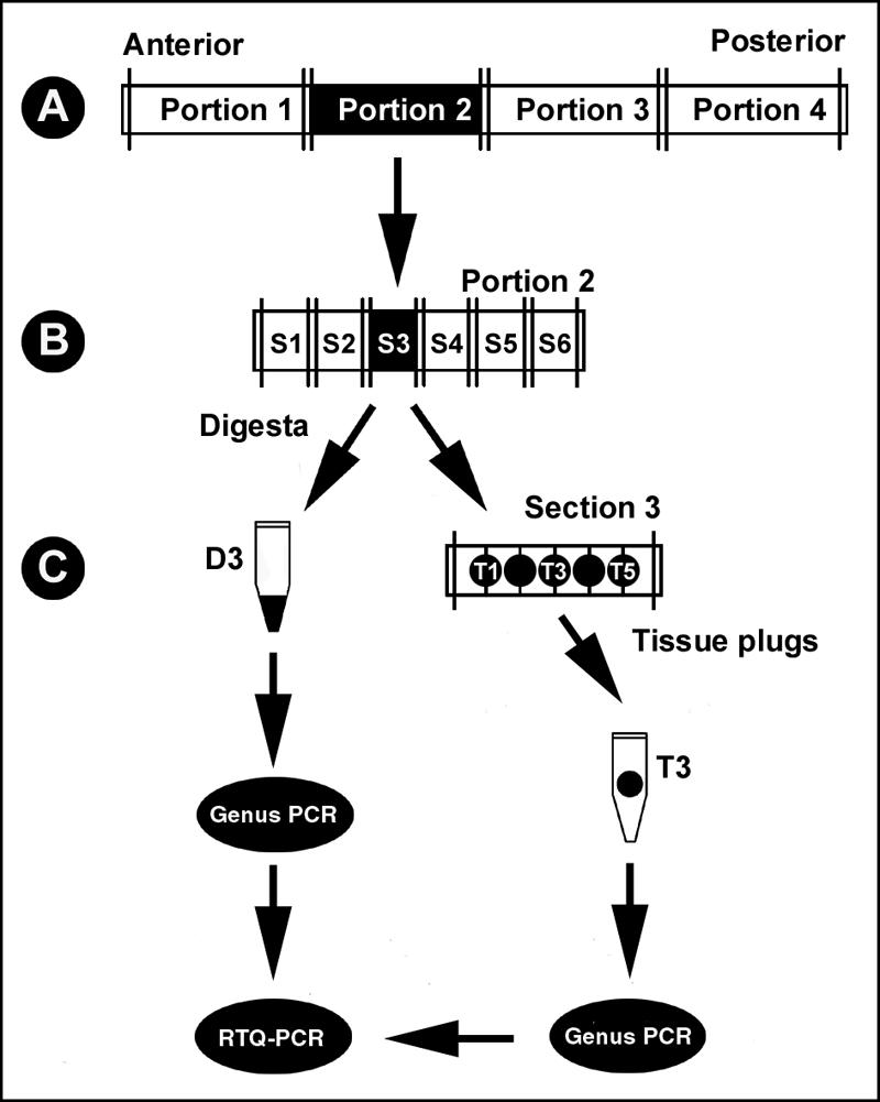
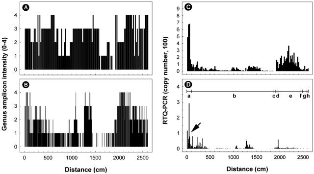
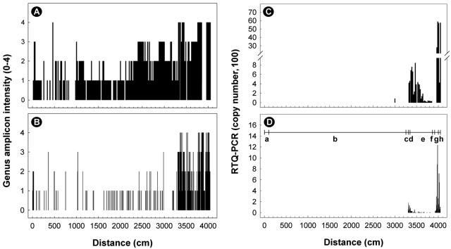
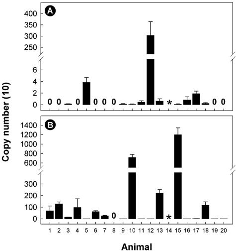
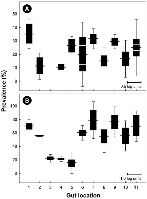
The location and abundance of Campylobacter jejuni and Campylobacter lanienae in the intestines of beef cattle were investigated using real-time quantitative PCR in two studies. In an initial study, digesta and tissue samples were obtained along the digestive tract of two beef steers known to shed C. jejuni and C. lanienae (steers A and B). At the time of slaughter, steer B weighed 540 kg, compared to 600 kg for steer A, yet the intestine of steer B (40.5 m) was 36% longer than the intestine of steer A (26.1 m). In total, 323 digesta samples (20-cm intervals) and 998 tissue samples (3.3- to 6.7-cm intervals) were processed. Campylobacter DNA was detected in the digesta and in association with tissues throughout the small and large intestines of both animals. Although C. jejuni and C. lanienae DNA were detected in both animals, only steer A contained substantial quantities of C. jejuni DNA. In both digesta and tissues of steer A, C. jejuni was present in the duodenum and jejunum. Considerable quantities of C. jejuni DNA also were observed in the digesta obtained from the cecum and ascending colon, but minimal DNA was associated with tissues of these regions. In contrast, steer B contained substantial quantities of C. lanienae DNA, and DNA of this bacterium was limited to the large intestine (i.e., the cecum, proximal ascending colon, descending colon, and rectum); the majority of tissue-associated C. lanienae DNA was present in the cecum, descending colon, and rectum. In a second study, the location and abundance of C. jejuni and C. lanienae DNA were confirmed in the intestines of 20 arbitrarily selected beef cattle. DNA of C. jejuni and C. lanienae were detected in the digesta of 57% and 95% of the animals, respectively. C. jejuni associated with intestinal tissues was most abundant in the duodenum, ileum, and rectum. However, one animal contributed disproportionately to the abundance of C. jejuni DNA in the ileum and rectum. C. lanienae was most abundant in the large intestine, and the highest density of DNA of this bacterium was found in the cecum. Therefore, C. jejuni colonized the proximal small intestine of asymptomatic beef cattle, whereas C. lanienae primarily resided in the cecum, descending colon, and rectum. This information could be instrumental in developing efficacious strategies to manage the release of these bacteria from the gastrointestinal tracts of cattle.


Figures

FIG. 1.
FIG. 1.
Schematic illustration of the method used for collection and processing of tissue and digesta samples from two chronically shedding beef cattle. (A) In the field, the digestive tract was divided into “portions” that were arbitrary lengths. (B) Subsequently, each portion was excised into 20-cm “sections” (designated S1, S2, S3, etc.). (C) Each section was longitudinally incised, and digesta was collected and frozen at −20°C. Following removal of digesta, tissue plugs (designated T1, T2, T3, etc.) were removed at 3.3-cm intervals, placed in microcentrifuge tubes, and frozen at −20°C. At designated locations, tissue plugs also were removed for microscopy. DNA was extracted from digesta and tissue plugs and subjected to conventional PCR for the genus Campylobacter and an internal amplification control and to real-time quantitative PCR to determine the numbers of C. jejuni and C. lanienae associated with digesta and each tissue plug.
FIG. 2.
FIG. 2.
Distribution of samples positive for Campylobacter DNA obtained from digesta (A and C) and tissues (B and D) of the intestinal tract of steer A. (A and B) Genus Campylobacter DNA examined with conventional PCR. The intensity of the genus amplicon was assessed based on a scale from 0 to 4 relative to a standard sample of known DNA. (C and D) Abundance of C. jejuni (genome copies in 2 μl of template) determined by nonnested real-time quantitative PCR targeting the mapA gene. The horizontal line with vertical lines in panel D indicates the various regions of the small and large intestines, where “a” is the duodenum, “b” is the jejunum, “c” is the ileum, “d” is the cecum, “e” is the ascending colon, “f” is the transverse colon, “g” is the descending colon, and “h” is the rectum. The arrow indicates a region where there was abundant C. jejuni DNA. The total length of the small and large intestines was 26.1 m.
FIG. 3.
FIG. 3.
Distribution of samples positive for Campylobacter DNA obtained from digesta (A and C) and tissues (B and D) of the intestinal tract of steer B. (A and B) Genus Campylobacter DNA examined with conventional PCR. The intensity of the genus amplicon was assessed based on a scale from 0 to 4 relative to a standard sample of known DNA. (C and D) Abundance of C. lanienae (genome copies in 2 μl of template) determined by using nested real-time quantitative PCR targeting the 16S rRNA gene. The horizontal line with vertical lines in panel D indicates the various regions of the small and large intestines, where “a” is the duodenum, “b” is the jejunum, “c” is the ileum, “d” is the cecum, “e” is the ascending colon, “f” is the transverse colon, “g” is the descending colon, and “h” is the rectum. The total length of the small and large intestines was 40.5 m.
FIG. 4.
FIG. 4.
Quantities of C. jejuni (A) and C. lanienae (B) DNA in digesta of 20 arbitrarily selected beef cattle. Quantities of DNA were determined using real-time quantitative PCR targeting the mapA (nonnested) and 16S rRNA (nested) genes for C. jejuni and C. lanienae, respectively. Most of the digesta samples were obtained from the rectum; the exceptions were animals 11 and 18, where samples were obtained from the descending colon. Neither an internal control amplicon nor an amplicon for the genus Campylobacter was obtained from the digesta sample indicated by the asterisk. The error bars indicate standard deviations (n = 2).
FIG. 5.
FIG. 5.
Prevalence of C. jejuni (A) and C. lanienae (B) associated with intestinal tissues of 20 beef cattle. The horizontal lines extending from the black bars indicate the percentages of positive animals (n = 20) for 11 locations in the small and large intestines. The locations are as follows: 1, proximal duodenum; 2, distal duodenum; 3, proximal jejunum; 4, central jejunum; 5, distal jejunum; 6, ileum; 7, free end of the cecum; 8, proximal loop of the ascending colon; 9, central flexure of the ascending colon; 10, descending colon; and 11, rectum. The solid bars indicate the relative abundance of each bacterium (mean log10 copy number in 2 μl of template). The vertical lines extending from the solid bars indicate standard deviations. The scale bars at the bottom right in the panels indicate mean template abundance (panel A, 0.5 log unit; panel B, 1.0 log unit). In panel A for gut locations 6 and 11, the areas delineated by the white lines in the bars indicate the mean values for C. jejuni template abundance minus the value for an animal with pneumonia.
FIG. 6.
FIG. 6.
Light micrographs showing the Campylobacter cells in association with mucosa of the distal duodenum of animal 13 obtained using the Hp Yellow and Hp Blue staining method (Anatech Ltd.). With this staining method, mucus appeared yellow, and bacteria within mucus and intestinal tissues appeared blue. (A) Crypt (Cr) with a layer of mucus stained yellow (Mu) coating the epithelium (Ep) stained blue with two Campylobacter cells also stained blue (arrows) associated with a strand of mucus (MuS). (B) Single Campylobacter cell (arrow; note the spiral morphology) associated with a thin strand of mucus within a duodenal crypt. Bars = 10 μm.

References

    1. Al-Mashat, R. R., and D. J. Taylor. 1980. Campylobacter spp. in enteric lesions in cattle. Vet. Rec. 107:31-34. - PubMed
    1. Al-Mashat, R. R., and D. J. Taylor. 1980. Production of diarrhoea and dysentery in experimental calves by feeding pure cultures of Campylobacter fetus subspecies jejuni. Vet. Rec. 107:459-464. - PubMed
    1. Al-Mashat, R. R., and D. J. Taylor. 1981. Production of enteritis in calves by the oral inoculation of pure cultures of Campylobacter fecalis. Vet. Rec. 109:97-101. - PubMed
    1. Al-Mashat, R. R., and D. J. Taylor. 1983. Bacteria in enteric lesions of cattle. Vet. Rec. 112:5-10. - PubMed
    1. Al-Mashat, R. R., and D. J. Taylor. 1983. Production of enteritis in calves by the oral inoculation of pure cultures of Campylobacter fetus subspecies intestinalis. Vet. Rec. 112:54-58. - PubMed

Publication types

MeSH terms

LinkOut - more resources