. 2005 Oct;115(10):2875-85.
doi: 10.1172/JCI25365. Epub 2005 Sep 8.

Apoptosis modulates protective immunity to the pathogenic fungus Histoplasma capsulatum

Affiliations

Apoptosis modulates protective immunity to the pathogenic fungus Histoplasma capsulatum

Holly L Allen et al. J Clin Invest. 2005 Oct.

Abstract

Pathogen-induced apoptosis of lymphocytes is associated with increased susceptibility to infection. In this study, we determined whether apoptosis influenced host resistance to the fungus Histoplasma capsulatum. The level of apoptotic leukocytes progressively increased in the lungs of naive and immune mice during the course of H. capsulatum infection. T cells constituted the dominant apoptotic population. Apoptosis was diminished in H. capsulatum-infected gld/gld and TNF-alpha-deficient mice; concomitantly, the fungal burden exceeded that of controls. Treatment of naive and H. capsulatum-immune mice with caspase inhibitors decreased apoptosis but markedly enhanced the severity of infection. Administration of a proapoptotic dose of suramin diminished the fungal burden. The increased burden in recipients of a caspase inhibitor was associated with elevations in IL-4 and IL-10 levels. In the absence of either of these cytokines, caspase inhibition suppressed apoptosis but did not increase the fungal burden. Thus, apoptosis is a critical element of protective immunity to H. capsulatum. Production of IL-4 and IL-10 is markedly elevated when apoptosis is inhibited, and the release of these cytokines exacerbates the severity of infection.


Figures

Figure 1
Figure 1
Apoptosis of lung leukocytes isolated from C57BL/6 mice infected with H. capsulatum. (A) In primary infection, mice were infected with 2 × 10⁶ yeast cells i.n. (B) In secondary infection, mice were infected with 104 yeast cells i.n. and 49 days later challenged with 2 × 10⁶ H. capsulatum yeast cells i.n. Apoptosis was assessed at 0, 7, 14, and 21 days after infection by flow cytometry. Data represent mean ± SEM of 6 animals per group. (C) Mice were infected with increasing numbers of H. capsulatum yeast cells (HC). Apoptosis was assessed at 7 days after infection. Data represent mean ± SEM of 6 animals per group. **P < 0.01 compared with each of the inocula. Data from 1 of 2 experiments are shown.
Figure 2
Figure 2
Phenotype of leukocytes isolated from lungs of mice infected with H. capsulatum for 7 days. Lung cells from mice with primary (A, C, and E) or secondary (B, D, and F) histoplasmosis were analyzed using allophycocyanin-labeled mAbs against the surface markers CD3ε, Mac1, or Gr1 (A and B) or PE-labeled mAbs against the surface markers CD4 and CD8 (C and D). Representative dot plots of at least 6 individual animals are shown. (E and F) Leukocytes were isolated at 7, 14, and 21 days after primary (E) and secondary (F) infection with histoplasmosis. The percentage of macrophages was calculated by subtracting the number of Gr1+ cells from the number of Mac1+ cells. Data represent mean ± SEM of 6 animals per group. Data from 1 of 2 experiments are shown.
Figure 3
Figure 3
Effect of TNF-α on apoptosis in mice infected with H. capsulatum. On the day of infection, mice were treated with either rat IgG or mAb against TNF-α in primary (A) or secondary (B) histoplasmosis. (C) The fungal burden was assessed for mice receiving mAb against TNF-α. (D) Wild-type C57BL/6 mice, TNFR1–/– mice, or TNFR2–/– mice were infected with 2 × 10⁶ yeast cells i.n. Data represent the mean ± SEM of 6 animals per group. *P < 0.05; **P = 0.01; #P < 0.001.
Figure 4
Figure 4
Apoptosis of lung leukocytes and fungal burden in gld/gld (GLD) mice. In primary infection (AD) and secondary infection (EH), lung leukocytes were analyzed for apoptosis (A, C, E, and G) or fungal burden assessed (B, D, F, and H). (C, D, G, and H) gld/gld mice were treated with either rat IgG or mAb to TNF-α. Data represent mean ± SEM of 6 animals per group. Data from 1 of 2 experiments are shown. **P < 0.01; #P < 0.001.
Figure 5
Figure 5
Effect of inhibitors of apoptosis on fungal burden in mice. Groups of mice were treated with either Boc-D-FMK (AF) or with suramin (G and H). Beginning the day of primary (AC) or secondary (DF) infection, mice were treated daily with HBSS, Boc-D-FMK, or Z-FA-FMK (10 μmol/kg). Lungs were harvested at 7 days after infection and were either assayed for apoptosis by flow cytometry (A and D) or evaluated for fungal burden (B and E). (C and F) Survival curves representing mice treated with either Boc-D-FMK or Z-FA-FMK daily until they succumbed to infection. Apoptosis (G) and fungal burden (H) were assessed in mice given z-DQMD-FMK. Data represent mean ± SEM of 6 animals per group. Pooled results from 2–3 experiments are shown. *P < 0.050; **P < 0.010; #P < 0.001.
Figure 6
Figure 6
The proapoptotic effect of suramin decreases fungal burden. Mice were treated with 1 mg of suramin i.p. or with HBSS alone at the time of infection. Lungs were harvested at 7 days postinfection and assayed for apoptosis by flow cytometry (A) and fungal burden (B). **P < 0.010.
Figure 7
Figure 7
Cytokine profiles of H. capsulatum–infected mice treated with the apoptosis inhibitor Boc-D-FMK. (AD) In primary infection, mice were infected with 2 × 10⁶ yeast cells i.n. (EH) In secondary infection mice were infected with 104 yeast cells i.n. and 49 days later challenged with 2 × 10⁶ H. capsulatum yeast cells i.n. Lungs were harvested at 7 days after infection, homogenized in HBSS, and assayed for TNF-α (A and E), IFN-γ (B and F), IL-10 (C and G), and IL-4 (D and H). Data represent the mean ± SEM of 6 animals per group. Data for 1 of 2 experiments are shown. *P < 0.050; **P < 0.010; #P < 0.001.
Figure 8
Figure 8
Apoptosis and fungal burden in IL-10–/– or IL-4–/– mice. Groups of IL-10–/– (A and B) or IL-4–/– (C and D) mice (n = 6) were infected with 2 × 10⁶ H. capsulatum yeast cells and administered PBS, Z-FA-FMK, or Boc-D-FMK for 1week. Lungs were analyzed for the proportion of apoptotic cells (A and C) and fungal burden (B and D). Data represent the mean ± SEM. **P < 0.010.
Figure 9
Figure 9
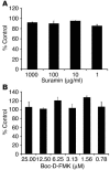
[3H]leucine incorporation by H. capsulatum yeast cells in the presence or absence of suramin (A) or Boc-D-FMK (B). The data are expressed as percent of control. For suramin, the control was yeast cells incubated with PBS. For Boc-D-FMK, the control was yeast cells incubated with Z-FA-FMK. Data represent the mean ± SEM of at least 5 experiments. Yeast cells were cultured in triplicate for each concentration of agent.

References

    1. Deepe, G.S., Jr. 2000. Histoplasma capsulatum. In Principles and practice of infectious diseases. 5th edition. G.L. Mandell, J.E. Bennett, and R. Dolin, editors. Churchill Livingstone. Philadelphia, Pennsylvania, USA. 2718–2732.
    1. Allendorfer R, Brunner GD, Deepe GS., Jr Complex requirements for nascent and memory immunity in pulmonary histoplasmosis. J. Immunol. 1999;162:7389–7396. - PubMed
    1. Deepe GS., Jr Role of CD8+ T cells in host resistance to systemic infection with Histoplasma capsulatum in mice. J. Immunol. 1994;152:3491–3500. - PubMed
    1. Gomez AM, Bullock WE, Taylor CL, Deepe GS., Jr Role of L3T4+ T cells in host defense against Histoplasma capsulatum. Infect. Immun. 1988;56:1685–1691. - PMC - PubMed
    1. Zhou P, Seder RA. CD40 ligand is not essential for induction of type 1 cytokine responses or protective immunity after primary or secondary infection with Histoplasma capsulatum. J. Exp. Med. 1998;187:1315–1324. - PMC - PubMed

Publication types

MeSH terms