An immunohistochemical study of the tissue bridging adult spondylolytic defects--the presence and significance of fibrocartilaginous entheses
- PMID: 16151708
- PMCID: PMC3489425
- DOI: 10.1007/s00586-005-0986-3
An immunohistochemical study of the tissue bridging adult spondylolytic defects--the presence and significance of fibrocartilaginous entheses
Abstract
Introduction Spondylolytic spondylolisthesis is an osseous discontinuity of the vertebral arch that predominantly affects the fifth lumbar vertebra. Biomechanical factors are closely related to the condition. An immunohistochemical investigation of lysis-zone tissue obtained from patients with isthmic spondylolisthesis was performed to determine the molecular composition of the lysis-zone tissue and enable interpretation of the mechanical demands to which the tissue is subject.
Methods: During surgery, the tissue filling the spondylytic defects was removed from 13 patients. Twelve spondylolistheses were at the L5/S1 level with slippage being less than Meyerding grade II. Samples were methanol fixed, decalcified and cryosectioned. Sections were labelled with a panel of monoclonal antibodies directed against collagens, glycosaminoglycans and proteoglycans.
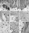
Results: The lysis-zone tissue had an ordered collagenous structure with distinct fibrocartilaginous entheses at both ends. Typically, these had zones of calcified and uncalcified fibrocartilage labelling strongly for type II collagen and aggrecan. Labelling was also detected around bony spurs that extended from the enthesis into the lysis-zone. The entheses also labelled for types I, III and VI collagens, chondroitin four and six sulfate, keratan and dermatan sulfate, link protein, versican and tenascin.
Conclusions: Although the gap filled by the lysis tissue is a pathological feature, the tissue itself has hallmarks of a normal ligament-i.e. fibrocartilaginous entheses at either end of an ordered collagenous fibre structure. The fibrocartilage is believed to dissipate stress concentration at the hard/soft tissue boundary. The widespread occurrence of molecules typical of cartilage in the attachment of the lysis tissue, suggests that compressive and shear forces are present to which the enthesis is adapted, in addition to the expected tensile forces across the spondylolysis. Such a combination of tensile, shear and compressive forces must operate whenever there is any opening or closing of the spondylolytic gap.
Figures


Similar articles
-
Fibrocartilage at the entheses of the suprascapular (superior transverse scapular) ligament of man--a ligament spanning two regions of a single bone.J Anat. 2001 Nov;199(Pt 5):539-45. doi: 10.1046/j.1469-7580.2001.19950539.x. J Anat. 2001. PMID: 11760885 Free PMC article.
-
The molecular composition of the extracellular matrix of the human iliolumbar ligament.Spine J. 2015 Jun 1;15(6):1325-31. doi: 10.1016/j.spinee.2013.07.483. Epub 2013 Oct 17. Spine J. 2015. PMID: 24139866
-
Molecular parameters indicating adaptation to mechanical stress in fibrous connective tissue.Adv Anat Embryol Cell Biol. 2005;178:1-71. Adv Anat Embryol Cell Biol. 2005. PMID: 16080262 Review.
-
Fibrocartilage in the transverse ligament of the human acetabulum.J Anat. 2001 Feb;198(Pt 2):223-8. doi: 10.1046/j.1469-7580.2001.19820223.x. J Anat. 2001. PMID: 11273046 Free PMC article.
-
Entheses--the bony attachments of tendons and ligaments.Ital J Anat Embryol. 2001;106(2 Suppl 1):151-7. Ital J Anat Embryol. 2001. PMID: 11729950 Review.
Cited by
-
Spontaneous pseudomeningocele associated with lumbar spondylolisthesis: A case report and review of the literature.Surg Neurol Int. 2017 Sep 7;8:221. doi: 10.4103/sni.sni_179_17. eCollection 2017. Surg Neurol Int. 2017. PMID: 28966827 Free PMC article.
-
An immunohistochemical study of the extracellular matrix of entheses associated with the human pisiform bone.J Anat. 2008 May;212(5):645-53. doi: 10.1111/j.1469-7580.2008.00887.x. Epub 2008 Apr 8. J Anat. 2008. PMID: 18399959 Free PMC article.
-
Is Preventative Long-Segment Surgery for Multi-Level Spondylolysis Necessary? A Finite Element Analysis Study.PLoS One. 2016 Feb 26;11(2):e0149707. doi: 10.1371/journal.pone.0149707. eCollection 2016. PLoS One. 2016. PMID: 26918333 Free PMC article.
References
-
- Bareggi R, Grill V, Zweyer M, Narducci P, Forabosco A. A quantitative study on the spatial and temporal ossification patterns of vertebral centra and neural arches and their relationship to the fetal age. Ann Anat. 1994;176:311–317. - PubMed
-
- Bono CM. Low-back pain in athletes. J Bone Joint Surg Am. 2004;86:382–396. - PubMed
Publication types
MeSH terms
Substances
Grants and funding
LinkOut - more resources
Full Text Sources
Miscellaneous

