Review
. 2005:59:451-85.
doi: 10.1146/annurev.micro.58.030603.123630.

Biogenesis, architecture, and function of bacterial type IV secretion systems

Affiliations
Review

Biogenesis, architecture, and function of bacterial type IV secretion systems

Peter J Christie et al. Annu Rev Microbiol. 2005.

Abstract

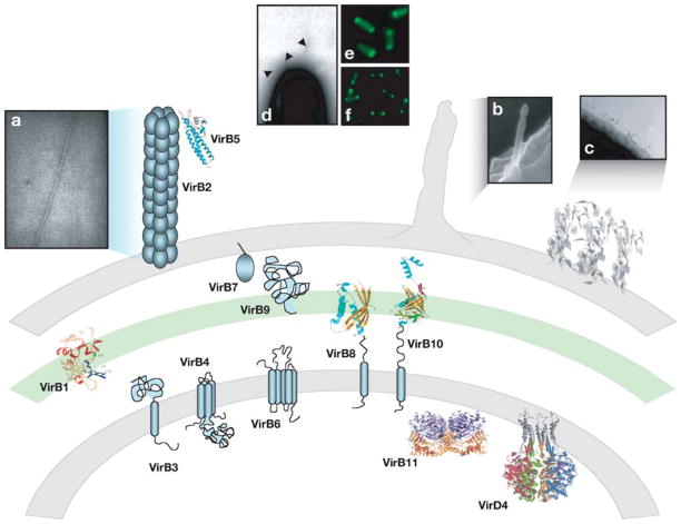
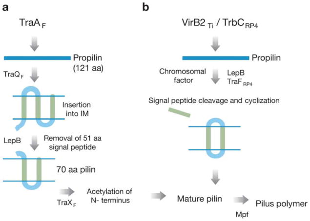
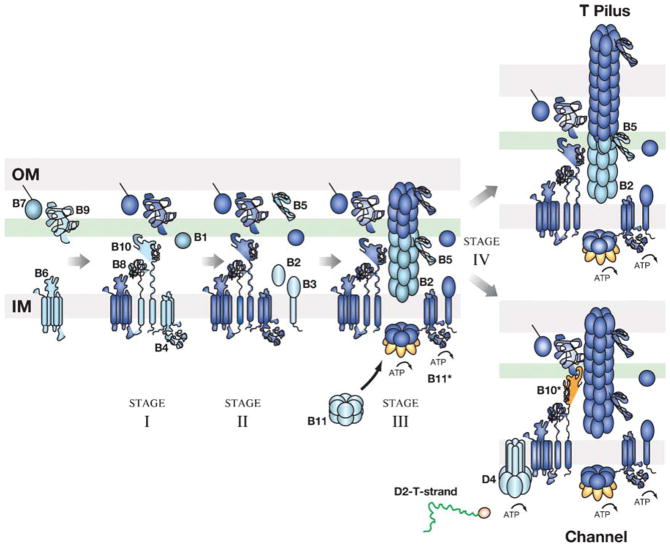
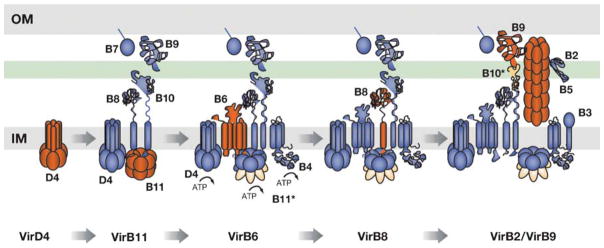
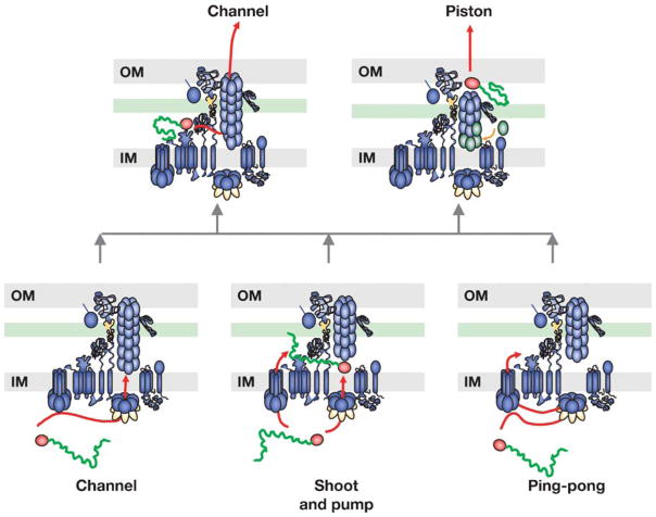
Type IV secretion (T4S) systems are ancestrally related to bacterial conjugation machines. These systems assemble as a translocation channel, and often also as a surface filament or protein adhesin, at the envelopes of Gram-negative and Gram-positive bacteria. These organelles mediate the transfer of DNA and protein substrates to phylogenetically diverse prokaryotic and eukaryotic target cells. Many basic features of T4S are known, including structures of machine subunits, steps of machine assembly, substrates and substrate recognition mechanisms, and cellular consequences of substrate translocation. A recent advancement also has enabled definition of the translocation route for a DNA substrate through a T4S system of a Gram-negative bacterium. This review emphasizes the dynamics of assembly and function of model conjugation systems and the Agrobacterium tumefaciens VirB/D4 T4S system. We also summarize salient features of the increasingly studied effector translocator systems of mammalian pathogens.


Figures

Figure 1
Figure 1
Topologies, structures, and cellular localizations of T4S subunits and organelles. Locations and structures or membrane topologies of the VirB/D4-like T4S subunits at the bacterial cell envelope. Microscopy images of (a) the A. tumefaciens T pilus, (b) H. pylori sheathed structure, and (c) L. pneumophila fibrous mesh are depicted. The A. tumefaciens VirB/D4 T4S system localizes at the cell poles as represented by positioning of (d) T pilus, (e) VirD4-GFP, and (f) VirB6-GFP. Three-dimensional structures of T4S subunits and surface organelles are reproduced with permission.
Figure 2
Figure 2
Processing of conjugative pilin monomers. Biochemical reactions leading to maturation of (a) TraAF and (b) VirB2Ti and TrbCRP4 pilin monomers and pilus formation are depicted. Pilin processing requires plasmid or chromosomal factors, and pilus polymerization requires the cognate Mpf proteins.
Figure 3
Figure 3
Biogenesis pathway of the A. tumefaciens VirB/D4 T4S system. A four-stage assembly pathway is presented for the A. tumefaciens VirB/D4 T4S system. Stage I: Assembly of the VirB6 plus B4-B7-B8-B9-B10 core complex; stage II: recruitment of B2-B3-B5 components; stage III: recruitment of hexameric VirB11 ATPase; stage IV: pathway bifurcation to yield the pilus or secretion channel. Subunits added at each stage of the pathway are depicted in blue-green, whereas subunits previously incorporated are depicted in dark blue. Conformational changes observed for the H. pylori HP0525 (B11*) N-terminal domain and A. tumefaciens VirB10 (B10*) are depicted in orange (*). We propose that progression through the stage IV reaction to form a functional secretion channel is regulated by intrinsic (ATP, substrate engagement) and extrinsic (recipient cell contact) signals. IM, inner membrane; OM, outer membrane.
Figure 4
Figure 4
Translocation pathway for the VirD2-T-strand transfer intermediate through the A. tumefaciens VirB/D4 secretion channel. The DNA substrate forms close contacts with VirB/D4 subunits in temporal order as delineated at the bottom. Subunits interacting with substrate at each step of the pathway are depicted in red, and subunits required at each successive subunit-substrate contact are depicted in blue. Substrate passage through the channel is depicted by a change in coloration in the channel subunit from red to blue. ATP energy is required for substrate transfer to VirB6 and each subsequent transfer step. Conformational changes in VirB11 and VirB10 required for substrate translocation are depicted in orange. IM, inner membrane; OM, outer membrane.
Figure 5
Figure 5
Proposed models for DNA substrate translocation across the A. tumefaciens cell envelope. The A. tumefaciens VirB/D4 T4S apparatus is represented as a transenvelope organelle that mediates passage of the nucleoprotein substrate (ball-line; red arrow). The different models proposed for substrate translocation through the inner membrane (bottom level: channel, shoot and pump, ping-pong) and through the outer membrane (top level: channel, piston) are depicted. Dynamic conformational changes (VirB11 ATP utilization; VirB2 pilin polymerization; VirB10 structural transition) required for substrate passage are highlighted in orange or green.

References

    1. Alegria MC, Docena C, Khater L, Ramos CHI, da Silva ACR, Farah CS. Identification of new protein-protein interactions involving products of the chromosome- and plasmid-encoded type IV secretion loci of the phytopathogen Xanthomonas axonopodis pv.citri. J Bacteriol. 2005;187:2315–25. - PMC - PubMed
    1. Amor JC, Swails J, Zhu X, Roy CR, Nagai H, et al. The structure of RalF, an ADP-ribosylation factor guanine nucleotide exchange factor from Legionella pneumophila, reveals the presence of a cap over the active site. J Biol Chem. 2005;280:1392–400. - PubMed
    1. Anderson LB, Hertzel AV, Das A. Agrobacterium tumefaciens VirB7 and VirB9 form a disulfide-linked protein complex. Proc Natl Acad Sci USA. 1996;93:8889–94. - PMC - PubMed
    1. Atmakuri K, Cascales E, Christie PJ. Energetic components VirD4, VirB11 and VirB4 mediate early DNA transfer reactions required for bacterial type IV secretion. Mol Microbiol. 2004;54:1199–211. - PMC - PubMed
    1. Atmakuri K, Ding Z, Christie PJ. VirE2, a type IV secretion substrate, interacts with the VirD4 transfer protein at cell poles of Agrobacterium tumefaciens. Mol Microbiol. 2003;49:1699–713. This study nicely integrates novel cytological and classical approaches to demonstrate that T4CP functions as a protein receptor for T4S machines. - PMC - PubMed

Publication types

MeSH terms

LinkOut - more resources