. 2005 Sep;13(9):1353-63.
doi: 10.1016/j.str.2005.06.006.

Mechanism of activation for transcription factor PhoB suggested by different modes of dimerization in the inactive and active states

Affiliations

Mechanism of activation for transcription factor PhoB suggested by different modes of dimerization in the inactive and active states

Priti Bachhawat et al. Structure. 2005 Sep.

Abstract

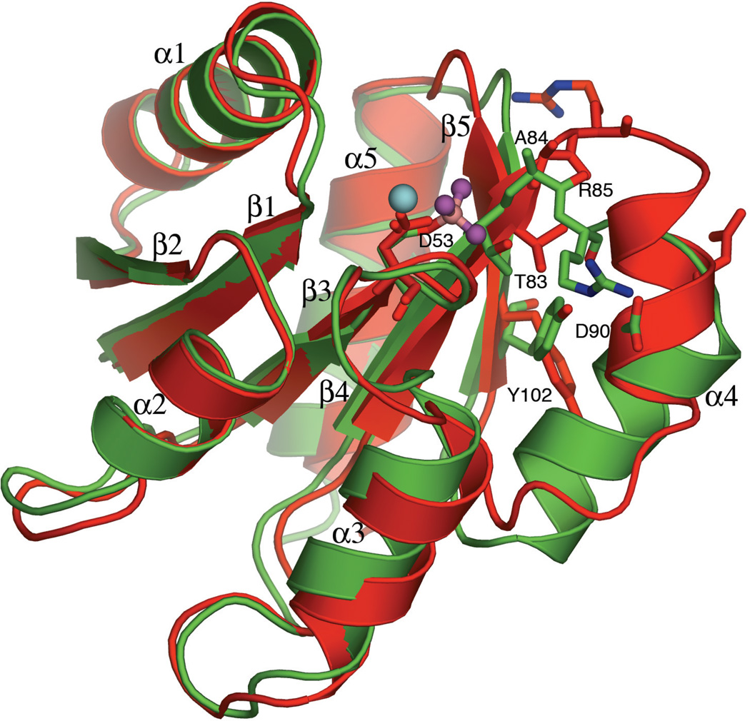
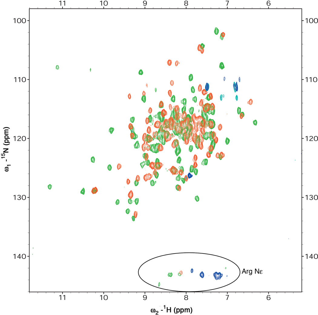
Response regulators (RRs), which undergo phosphorylation/dephosphorylation at aspartate residues, are highly prevalent in bacterial signal transduction. RRs typically contain an N-terminal receiver domain that regulates the activities of a C-terminal DNA binding domain in a phosphorylation-dependent manner. We present crystallography and solution NMR data for the receiver domain of Escherichia coli PhoB which show distinct 2-fold symmetric dimers in the inactive and active states. These structures, together with the previously determined structure of the C-terminal domain of PhoB bound to DNA, define the conformation of the active transcription factor and provide a model for the mechanism of activation in the OmpR/PhoB subfamily, the largest group of RRs. In the active state, the receiver domains dimerize with 2-fold rotational symmetry using their alpha4-beta5-alpha5 faces, while the effector domains bind to DNA direct repeats with tandem symmetry, implying a loss of intramolecular interactions.


Figures

Figure 1
Figure 1
Crystal Structures Show Different Dimers for Inactive and Active Regulatory Domains of PhoB. (A) Inactive PhoBN dimerizes using the α1–α5 interface. Inactive PhoBN is red; the dimer interface is beige. (B) Active PhoBN dimerizes using the α4-β5-α5 interface. Active PhoBN is green; the dimer interface is blue; and active site ligands are shown as spheres with magnesium in cyan, beryllium in pink, and fluorides in magenta. Figures were created using Pymol (http://pymol.sourceforge.net/).
Figure 2
Figure 2
HN-15N HSQC Spectra of Inactive and Active Regulatory Domains of PhoB. The number of peaks observed in the HSQC spectra are 124 and 128 for inactive (red) and active (green) PhoBN, respectively, both corresponding to monomers or two-fold symmetric dimers. At NMR concentrations, both proteins exist as symmetric dimers (see text). The large differences in the spectra of inactive and active PhoBN suggest that the structures of the two dimers are significantly different.
Figure 3
Figure 3
Dilution Experiments of Inactive PhoBN. Successive dilutions of inactive PhoBN from 1.5 mM to 0.1 mM cause a progressive shift in some peaks indicating a fast monomer-dimer equilibrium on the NMR timescale (i.e. τ ≪ 100 ms). A section of the TROSY-HSQC spectrum is shown, with the inset zoomed on one of the resonances progressively shifting with decreasing concentration. Crosspeaks for 1.5 mM protein are shown in blue, for 0.75 mM in green, for 0.375 mM in yellow, and for 0.1 mM in red.
Figure 4
Figure 4
The Active Site of BeF3-activated PhoBN. A stereo view of a difference electron density map (Fobs-Fcalc) calculated at 2 Å with omission of the Mg2+, BeF3 and active site water molecules from the model is shown in blue, contoured at 3σ. Waters and the catalytic Mg2+ are shown as red and cyan spheres, respectively. The protein and BeF3 complex are shown in stick representation; beryllium is pink and fluorides are magenta (associations within the non-covalent BeF3 complex are shown as bonds for clarity).
Figure 5
Figure 5
Conformational Changes in PhoBN upon Activation. The greatest conformational changes upon activation occur in the β4–α4 loop, the α4 helix and the α5-β5 loop. The important switch residues Thr83 and Tyr102, residues Ala84 and Arg85 in the β4–α4 loop, residue Asp90 in helix α4 and active site residue Asp53 are shown in stick representation. The inactive protein is red and the active protein is green. Active site ligands are shown in ball-and-stick representation; Mg2+ is cyan, beryllium is pink and fluorides are magenta.
Figure 6
Figure 6
Stereo View of the α4-β5-α5 Interface of Active PhoBN. The residues involved in salt bridge and hydrophobic interactions at the interface are completely conserved in, and only in, the OmpR/PhoB subfamily of RRs. Residues involved in salt bridges are shown as sticks, and the hydrophobic residues are shown as spheres. The two protomers of the dimer are colored orange and green. Salt bridges are shown as dotted lines.
Figure 7
Figure 7
HetNOE Spectra of Inactive and Active PhoBN. Negative resonances for Hof arginines in the spectrum of inactive PhoBN, indicating disordered arginines, become positive upon activation, indicating order, possibly due to involvement in salt bridges at the dimer interface of the active protein. (A) For inactive PhoBN, positive resonances are colored red and negative resonances are colored blue; for active PhoBN, positive resonances are colored green and no negative resonances are observed. (B) 1D slices through the arginine H region of the HetNOE spectrum of inactive PhoBN show positive and negative resonances (top), whereas for activated PhoBN, all arginine H resonances are positive (bottom).
Figure 8
Figure 8
Model of Phosphorylation-Induced Activation in the OmpR/PhoB Subfamily. In the active state, believed to be common to all OmpR/PhoB subfamily members, the receiver domains form a two-fold symmetric dimer while the DNA-binding domains bind to DNA with tandem symmetry. Inactive PhoB exists in equilibrium between a monomer and dimer. The inactive α1–α5 dimer, specific to PhoB, provides an additional means of inhibition by positioning the effector domains in opposite directions, incompatible with tandem DNA binding. Inactive PhoBN is shown in red, active PhoBN in green, the α1–α5 interface of the inactive dimer in beige, the α4-β5–α5 interface of the active dimer in blue, the DNA-binding domain in grey, and the recognition helix in pink. Full-length inactive PhoB was modeled by positioning the two domains of PhoB as they exist in the structure of DrrB.

Comment in

References

    1. Aiba H, Mizuno T. Phosphorylation of a bacterial activator protein, OmpR, by a protein kinase, EnvZ, stimulates the transcription of the ompF and ompC genes in Escherichia coli. FEBS Lett. 1990;261:19–22. - PubMed
    1. Aoyama T, Takanami M, Makino K, Oka A. Cross-talk between the virulence and phosphate regulons of Agrobacterium tumefaciens caused by an unusual interaction of the transcriptional activator with a regulatory DNA element. Mol. Gen. Genet. 1991;227:385–390. - PubMed
    1. Baikalov I, Schröder I, Kaczor-Grzeskowiak M, Grzeskowiak K, Gunsalus RP, Dickerson RE. Structure of the Escherichia coli response regulator NarL. Biochemistry. 1996;35:11053–11061. - PubMed
    1. Bellsolell L, Prieto J, Serrano L, Coll M. Magnesium binding to the bacterial chemotaxis protein CheY results in large conformational changes involving its functional surface. J. Mol. Biol. 1994;238:489–495. - PubMed
    1. Bent CJ, Isaacs NW, Mitchell TJ, Riboldi-Tunnicliffe A. Crystal structure of the response regulator 02 receiver domain, the essential YycF two-component system of Streptococcus pneumoniae in both complexed and native states. J. Bacteriol. 2004;186:2872–2879. - PMC - PubMed

Publication types

MeSH terms

Associated data

LinkOut - more resources