. 2005 Aug 23;5(1):89-94.
doi: 10.1102/1470-7330.2005.0019.

Retroperitoneal sarcomas

Affiliations

Retroperitoneal sarcomas

Isaac R Francis et al. Cancer Imaging. .

Abstract

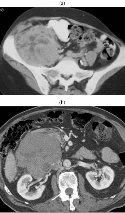
Retroperitoneal sarcomas are rare neoplasms. CT or MR imaging is performed in patients with these tumors to detect local extent and distant metastases of the tumor and for preoperative surgical planning. Most sarcomas cannot be characterized as to cell type with CT or MR, with the exceptions being liposarcomas and intracaval leiomyosarcomas. Similarly histological grading cannot be made definitively with imaging alone, the exception being liposarcoma since well differentiated liposarcomas contain more macroscopic fat than do less differentiated liposarcomas. After surgery, follow up imaging with CT or MR and careful scrutiny of the tumor bed and resection site are essential to detect early recurrences, which can often be managed with re-resection.


Figures

Figure 1
Figure 1
Contrast-enhanced axial CT shows two large retroperitoneal heterogenous neoplasms with areas of low density due to degeneration or necrosis. These proved to be malignant: a peripheral nerve sheath tumor (a) and retroperitoneal leiomyosarcoma (b), respectively.
Figure 2
Figure 2
Liposarcoma. Contrast-enhanced axial CT shows large right retroperitoneal liposarcoma (arrow) composed predominantly of fat but also has areas of soft tissue density and calcific components.
Figure 3
Figure 3
High-grade liposarcoma. Contrast-enhanced axial CT shows left-sided predominantly soft-tissue density abdominal tumor (arrow) which proved to be a high-grade pleomorphic liposarcoma.
Figure 4
Figure 4
Intracaval leiomyosarcoma. Large inferior vena caval leiomyosarcoma (arrow) seen on (a) contrast-enhanced CT and (b) coronal contrast-enhanced gradient echo MR image (arrow). Note intraluminal tumor extension on both images.
Figure 5
Figure 5
Well-differentiated low-grade and high-grade liposarcomas. (a) Contrast-enhanced axial CT shows well encapsulated fatty mass (arrow) with no septations or soft tissue component. This proved to be a well-differentiated liposarcoma. (b) Contrast-enhanced axial CT shows a predominantly soft tissue pelvic mass (arrow) which proved to be a high-grade liposarcoma.
Figure 6
Figure 6
Metastatic fibrosarcoma. (a) Coronal PET-FDG image shows peritoneal metastatic nodule (arrow). (b) Axial contrast-enhanced CT shows this metastatic nodule (arrow), which was not prospectively identified as it was thought to be part of the left lobe of the liver.
Figure 7
Figure 7
Recurrent liposarcoma. (a) Contrast-enhanced axial CT shows right retroperitoneal mass (arrow) which as a liposarcoma. (b) On a 6-month post-resection follow-up contrast-enhanced axial CT, there is suggestion of recurrence (arrow). This was interpreted as being indeterminate for recurrence. (c) At 12-month follow-up contrast-enhanced axial CT, the mass (arrow) has shown interval growth and is more obvious. This proved to be recurrent liposarcoma.

References

    1. Mettlin C, Priore R, Rao U. Results of the national soft tissue sarcoma registry. J Surg Oncol. 1982;19:224–7. - PubMed
    1. McGrath P. Retroperitoneal sarcomas. Semin Surg Oncol. 1994;10:364–8. - PubMed
    1. Daugaard S. Current soft tissue sarcoma classification. Eur J Cancer. 2004;40:543–8. - PubMed
    1. Coindre JM, Mariani O, Chibon F, et al. Most malignant fibrous histiocytomas developed in the retroperitoneum are dedifferentiated liposarcomas: a review of 25 cases initially diagnosed as malignant fibrous histiocytomas. Mod Pathol. 2003;16:256–62. - PubMed
    1. Papanicolaou N, Yoder IC, Lee MJ. Primary retroperitoneal neoplasms: How close can we come in making the correct diagnosis. Urol Radiol. 1992;14:221–8. - PubMed

LinkOut - more resources