. 2005 Sep;26(8):1986-95.

West Nile virus meningoencephalitis: MR imaging findings

Affiliations

West Nile virus meningoencephalitis: MR imaging findings

Kalliopi A Petropoulou et al. AJNR Am J Neuroradiol. 2005 Sep.

Abstract

Background and purpose: Reports of MR imaging in West Nile virus (WNV) meningoencephalomyelitis are few and the described findings limited. The purpose of this study was to review the spectrum of MR imaging findings for WNV meningoencephalomyelitis and investigate whether any of the findings correlates with clinical presentation of flaccid paralysis.

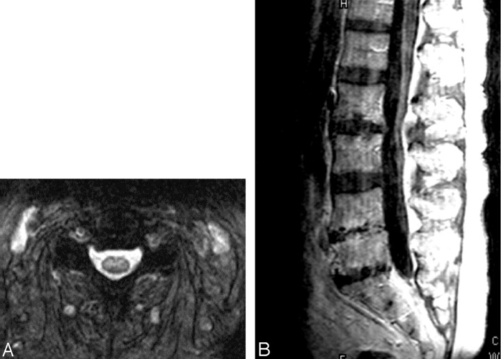
Methods: We reviewed the MR imaging findings of 17 patients with confirmed WNV encephalitis and/or myelitis. MR imaging brain studies were evaluated for location of signal intensity abnormalities, edema, hydrocephalus, or abnormal enhancement. MR imaging spine studies were evaluated for signal intensity abnormalities in cord and/or enhancement.

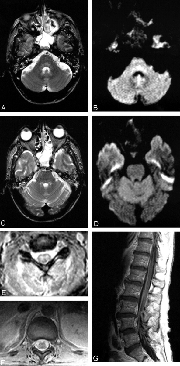
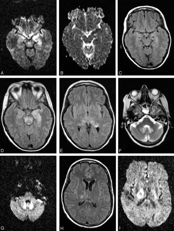
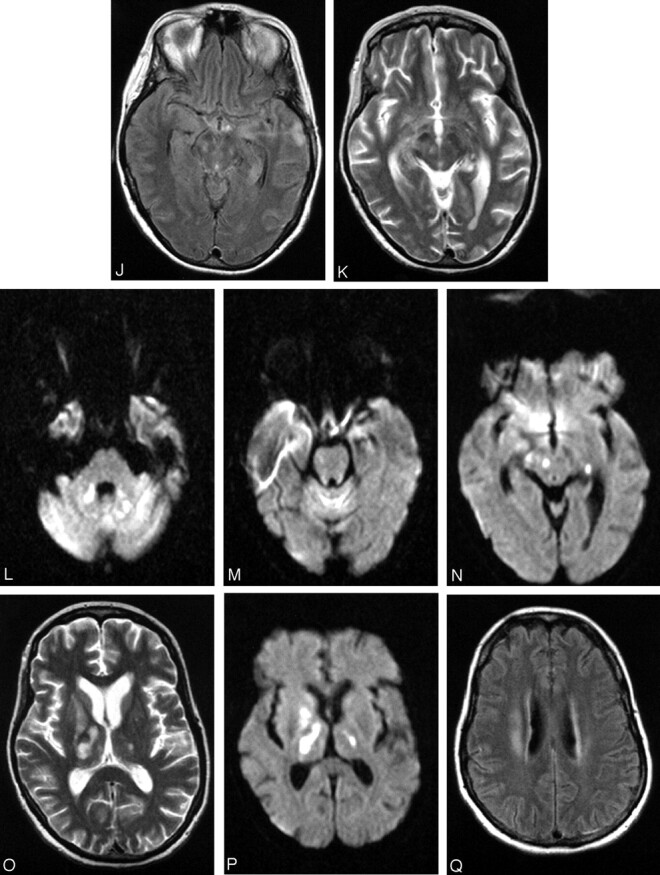
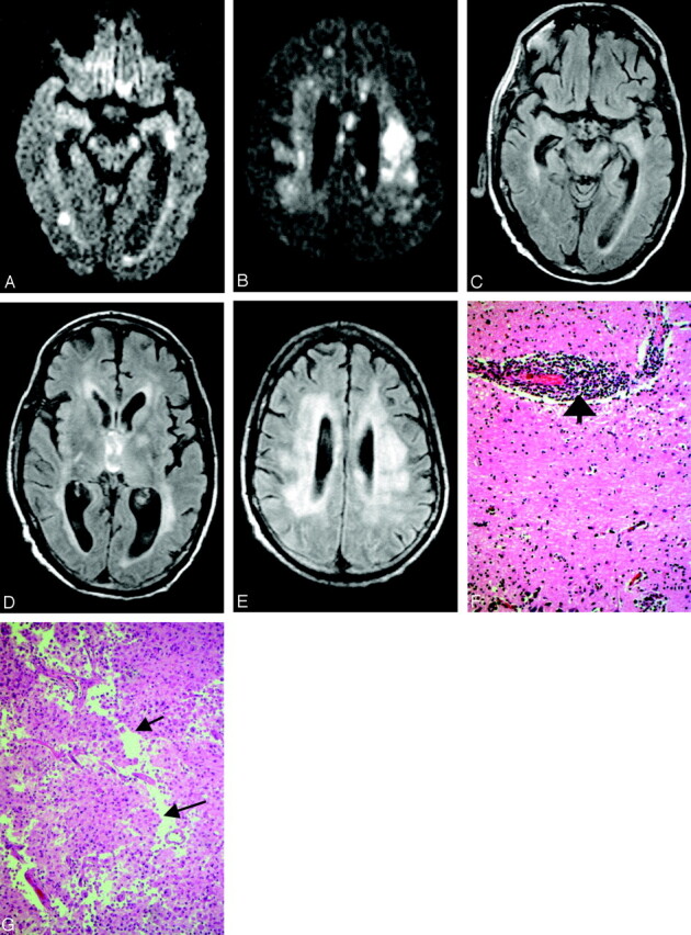
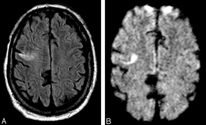
Results: Retrospective review of the MR imaging studies of 17 patients was performed by 2 neuroradiologists. Eleven of 16 brain MR images demonstrated abnormalities. Eight (50%) patients had abnormal studies related to meningoencephalitis. All 8 patients had abnormal findings in the deep gray matter and/or brain stem; 2 had additional white matter abnormalities. Three patients with abnormal MR studies of the spine had extremity weakness on examination. The imaging findings included abnormal signal intensity more pronounced in the ventral horns and/or enhancement around the conus medullaris and cauda equina. One patient had additional abnormalities in the pons.

Conclusion: Abnormal MR imaging findings in patients with WNV meningoencephalomyelitis are nonspecific but not uncommon. Anatomic areas commonly affected are basal ganglia, thalami, mesial temporal structures, brain stem, and cerebellum. Extremity weakness or flaccid paralysis corresponds to spinal cord/cauda equina abnormalities.


Figures

F<sc>ig</sc> 1.
Fig 1.
Patient 2, a 40-year-old man admitted with encephalitis and developed flaccid paralysis during hospitalization: axial FSE T2-weighted (A) and trace DW (B) images demonstrate increased signal intensity in the pontine tegmentum. Increased signal intensity was also seen in the superior cerebellar peduncles on FSE T2-weighted image (C), but not on the trace DW image (D). The patient was intubated at the time of imaging, which resulted in opacified sinuses and mastoid air cells. Axial GRE sequences through the cervical (E) and thoracic cord (F) show abnormal signal intensity in the gray matter with more pronounced involvement of the ventral horns. Postcontrast sagittal T1-weighted image (G) demonstrates enhancement of the cauda equina.
F<sc>ig</sc> 2.
Fig 2.
Patient 17, a 49-year-old woman with history of non-Hodgkin lymphoma in remission presented with fever. Work-up was negative for recurrence. The first MR imaging of the brain shows abnormality in the left mesial temporal lobe on trace DW image (A) and ADC (B), though the finding is subtle on the FLAIR sequences (C). At the time of imaging, the findings raised the question of herpes encephalitis, for which the patient was originally treated. Following deterioration of the mental status and development of upper extremity weakness, a second MR imaging was obtained. The abnormality is now apparent on the FLAIR sequences and has progressed to involve not only the contralateral mesial temporal structures, but also the substantia nigra (D), as well as the mesial and dorsal aspect of the thalami (E). Further clinical deterioration with established “polio-like” symptoms prompted a new MR imaging 11 days later, which demonstrated new areas of involvement with resolution of improvement of prior lesions. Images of this MR imaging show increased signal intensity in the dentate nuclei on FLAIR (F) and DW (G) images and right thalamus on FLAIR image (2 hours) and DW image (I) and improvement of the mesial temporal lobe and midbrain abnormalities (J). Focus of T2 hyperintensity is seen in the right red nucleus on FSE T2-weighted image (K). On J, note site of biopsy in the lateral aspect of the left temporal lobe that was negative for herpes encephalitis. The fourth MR imaging was obtained upon patient’s discharge to a nursing facility. DW images show increased signal intensity in the cerebellar hemispheres and right branchium pontis (L), vermis (M), and the right red nucleus (N). Cerebellar hemisphere abnormalities were more conspicuous on DW than FLAIR or FSE T2-weighted images. FSE T2-weighted (O) and DW (P) images through the level of the basal ganglia demonstrate persistent abnormality in the right thalamus and new lesions in the right globus pallidus and left thalamus, whereas the FLAIR image demonstrates involvement of the right corona radiata (Q) as well.
F<sc>ig</sc> 3.
Fig 3.
Patient 11, a 51-year-old man post renal and pancreatic transplant with recent mosquito exposure. Axial DW images show patchy areas of increased signal intensity in the periventricular white matter and left cerebral peduncle (A) as well as corona radiata and corpus callosum (B). Axial FLAIR sequences demonstrate subtle abnormalities in the left mesial temporal lobe, both cerebral peduncles, more pronounced in the left one (C), and both thalami and left globus pallidus (D). Diffusely increased signal intensity is also present in the deep hemispheric white matter (D and E). Histopathology (F) shows the hippocampus with marked perivascular (bold arrow) lymphocytic inflammation (hematoxylin and eosin; original magnification 200×). Histopathology (G) demonstrates white matter infarct with necrosis, macrophage and early cavitary changes (cavitation delineated by arrows) (hematoxylin and eosin; original magnification 200×).
F<sc>ig</sc> 4.
Fig 4.
Patient 16, an 80-year-old man presented with extremity weakness. Axial GRE image through cervical spine (A) shows increased signal intensity in the gray matter, whereas postcontrast sagittal T1-weighted sequences (B) reveal nerve root enhancement.
F<sc>ig</sc> 5.
Fig 5.
Patient 1, a 75-year-old man showed increased signal intensity in the right frontal lobe (A) with corresponding abnormality on the DW image (B), which was prospectively interpreted as infarct.

References

    1. Nash D, Mostashari F, Fine A, et al. The outbreak of West Nile virus infection in New York City area in 1999. N Engl J Med 2001;344:1807–1814 - PubMed
    1. Jeha LE, Sila CA, Lederman RJ, et al. West Nile virus infection: a new acute paralytic illness. Neurology 2003;61:55–59 - PubMed
    1. Olsan A, Milburn JM, Baumgarten KL, Durham HL. Leptomeningeal enhancement in a patient with proven West Nile virus infection. AJR Am J Roentgenol 2003;181:591–592 - PubMed
    1. Rosas H, Wippold II FJ. West Nile virus: case report with MR imaging findings. AJNR Am J Neuroradiol 24:1376–1378 - PMC - PubMed
    1. Lanciotti RS, Roehrig JT, Deubel V, et al. Origin of the West Nile virus responsible for an outbreak of encephalitis in the northeastern United States. Science 1999;286:2333–2337 - PubMed