

Case Reports
The hypodense artery sign
Affiliations
- PMID: 16155153
- PMCID: PMC8148856
Item in Clipboard
Case Reports
The hypodense artery sign
AJNR Am J Neuroradiol.
2005 Sep.
Abstract
An acute intracranial embolus may be associated with a hyperdense artery sign on CT, related to acute thrombus within the affected artery, a phenomenon that is well known and has been extensively documented. We present an unusual case of a middle cerebral artery territory acute infarct with a hypodense artery on CT. The Hounsfield unit attenuation of the embolic lesion was fat density. CT angiography and MR imaging confirmed the fatty lesion to be within the middle cerebral artery.
Figures
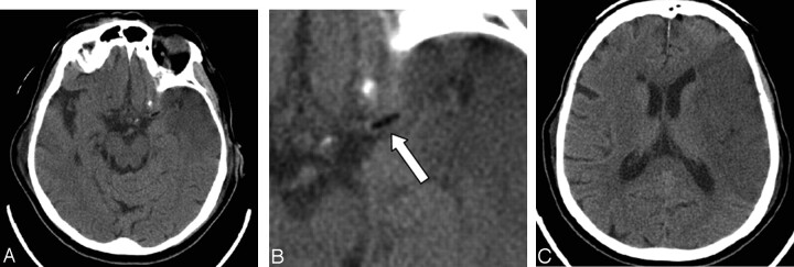
A 78-year-old woman who had undergone a mitral valve replacement. A–C, Axial images from the initial CT head scan obtained on the first postoperative day demonstrate an early left middle cerebral artery territory infarct. There is a fat attenuation filling defect (arrow, B) in the region of the proximal left middle cerebral artery trunk.
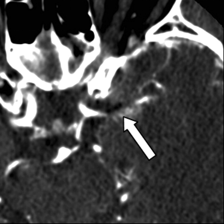
CT angiography axial image obtained 13 days postoperatively confirms that the fat attenuation lesion (arrow) is within the proximal left middle cerebral artery trunk.
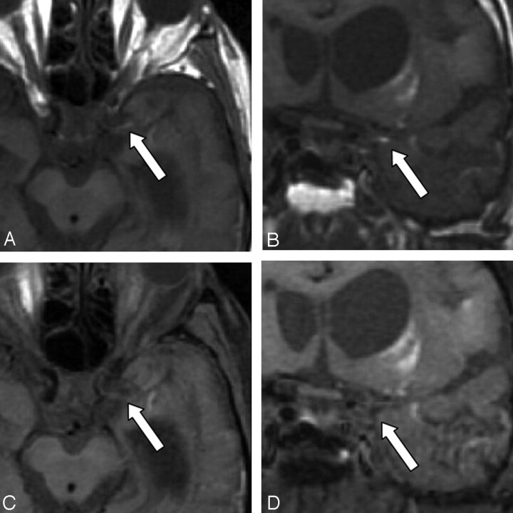
Axial and coronal T1-weighted MR images obtained 49 days postoperatively without fat saturation (A,B) and with fat saturation (C,D). The images without fat saturation demonstrate the T1-hyperintense embolus (arrow) in the proximal left M1 trunk. After fat saturation, the T1 hyperintensity of the embolus is saturated (arrow), confirming its fatty composition. High-signal-intensity methemoglobin in the adjacent infarct is unchanged before and after fat saturation.
References
-
- von Kummer R, Nolte PN, Schnittger H, Thron A, Ringelstein EB. Detectability of cerebral hemisphere ischaemic infarction by CT within 6 hours of stroke. Neuroradiology 1996;38:31–33 - PubMed
-
- Gacs G, Fox AJ, Barnett HJM, Vinuela F. CT visualization of intracranial arterial thromboembolism. Stroke 1983;14:756–762 - PubMed
-
- Pressman BD, Tourje EJ, Thompson JR. An early sign of ischemic infarction: increased density in a cerebral artery. AJNR Am J Neuroradiol 1987;8:645–648 - PubMed
-
- Schuierer G, Huk W. The unilateral hyperdense middle cerebral artery: an early CT sign of embolism or thrombosis. Neuroradiology 1988;30:120–122 - PubMed
-
- Bastianello S, Pierallini A, Colonnese C, et al. Hyperdense middle cerebral artery CT sign: comparison with angiography in the acute phase of ischemic supratentorial infarction. Neuroradiology 1991;33:207–211 - PubMed
Publication types
MeSH terms
LinkOut - more resources
Full Text Sources
Medical