

Review
Primary intraosseous meningioma: CT and MRI appearance
Affiliations
- PMID: 16155159
- PMCID: PMC8148822
Item in Clipboard
Review
Primary intraosseous meningioma: CT and MRI appearance
AJNR Am J Neuroradiol.
2005 Sep.
Abstract
Benign primary intraosseous meningioma presenting with osteolytic skull lesion and soft-tissue component is rare. CT and MR imaging of a patient with frontoparietal scalp swelling showed an osteolytic intracalvarial lesion with an extradural soft-tissue component. Following wide surgical resection, the histological examination revealed an intraosseous chordoid meningioma. The clinical and radiological findings of primary intraosseous meningioma are discussed and the relevant literature is reviewed.
Figures
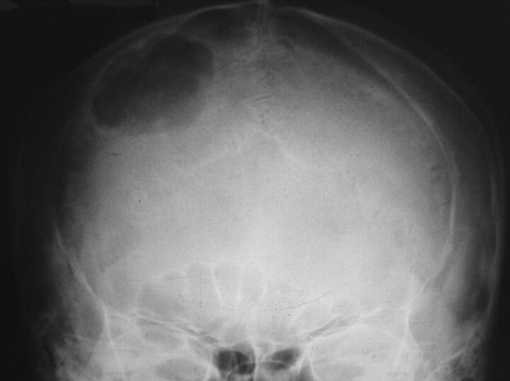
Plain skull radiograph shows a well-defined solitary lytic lesion in the right frontoparietal region.
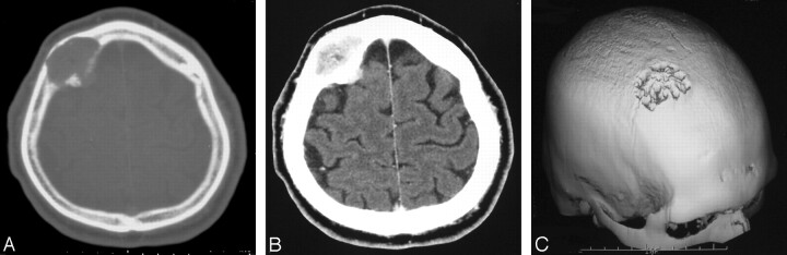
CT scan with bone window (A) demonstrates a right-sided frontoparietal intradiploic mass expanding the calvaria with cortical destruction. Postcontrast CT scan (B) shows prominent enhancement and intracranial extension of the lesion. Three-dimensional reformatted CT image (C) clearly demonstrates the osteolytic skull lesion.
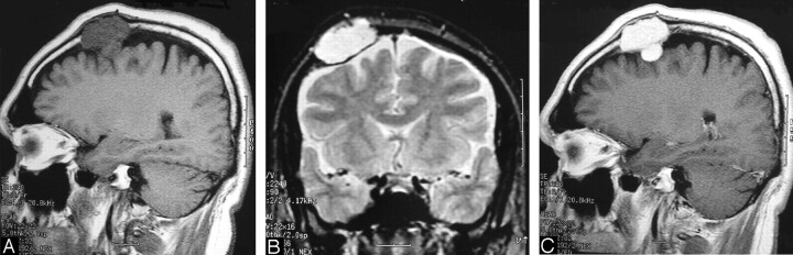
Sagittal T1-weighted (A) and coronal T2-weighted (B) MR images show the frontoparietal intracalvarial mass lesion that was hypointense on T1-(A) and hyperintense on T2-weighted (B) images. The lesion shows intense and homogeneous enhancement on postcontrast T1-weighted (C) image. MR images (A–C) reveal the intracranial extension and extradural location of the lesion.
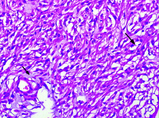
Photomicrograph of the tumor shows the chordoid meningioma with eosinophilic vacuolated tumor cells (large arrow) in a mucous-rich matrix (small arrow) (hematoxylin and eosin, original magnification ×200).
References
-
- Muzumdar DP, Vengsarkar US, Bhatjiwale MG, Goel A. Diffuse calvarial meningioma: a case report. J Postgrad Med 2001;47:116–118 - PubMed
-
- Whicker JH, Devine KD, McCarty CS. Diagnostic and therapeutic problems in extracranial meningiomas. Am J Surg 1973;123:452–457 - PubMed
-
- Lang FF, Macdonald OK, Fuller GN, DeMonte F. Primary extradural meningiomas: a report on nine cases and review of literature from the era of computerized tomography scanning. J Neurosurg 2000;93:940–950 - PubMed
-
- Hoye SJ, Hoar CS, Murray JE. Extracranial meningioma presenting as a tumor of the neck. Am J Surg 1960;100:486–489 - PubMed
-
- Crawford TS, Kleinschmidt-Demasters BK, Lillehei KO. Primary intraosseous meningioma: case report. J Neurosurg 1995;83:912–915 - PubMed
Publication types
MeSH terms
LinkOut - more resources
Full Text Sources
Medical