. 2005 Sep;26(8):2086-94.

Identification, prognosis, and management of patients with carotid artery near occlusion

Affiliations

Identification, prognosis, and management of patients with carotid artery near occlusion

Allan J Fox et al. AJNR Am J Neuroradiol. 2005 Sep.

Abstract

Background and purpose: Two large trials indicated that endarterectomy was less beneficial for symptomatic patients with internal carotid artery (ICA) near occlusion than for patients who had severe stenosis without near occlusion. Near occlusions complicate ratio calculations of ICA stenosis and require attention to detail for identification. The goal is to provide diagnostic criteria, illustrate identifying features, estimate accuracy of identification, and assess prognosis for patients with near occlusion.

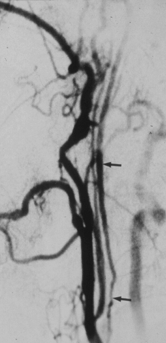
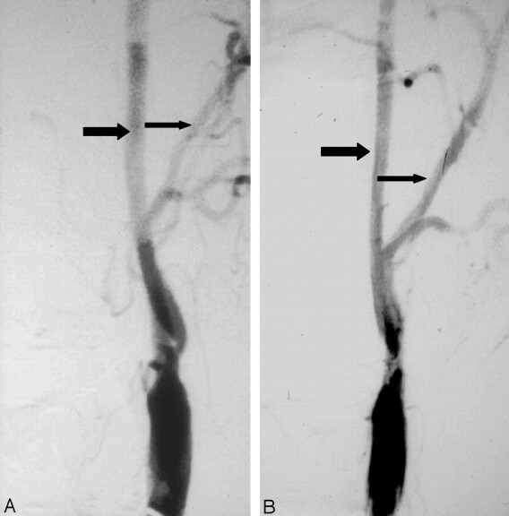
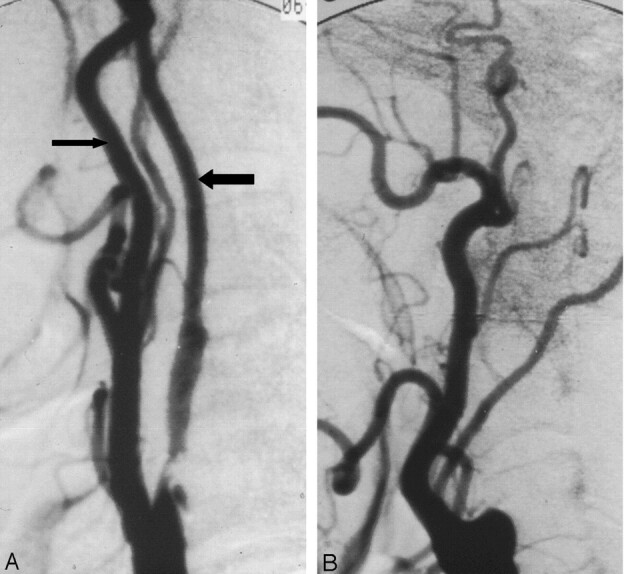
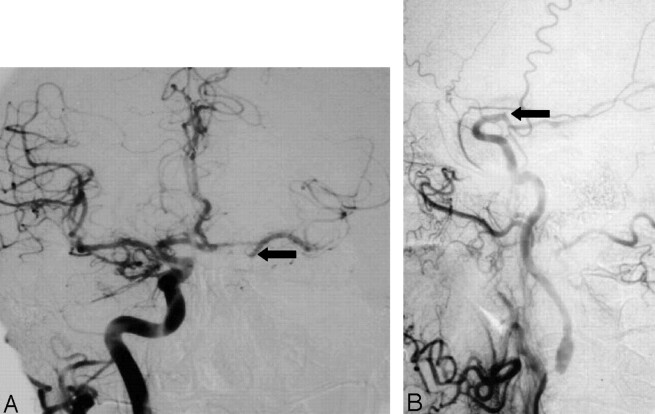
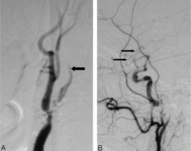
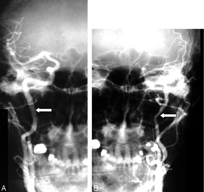
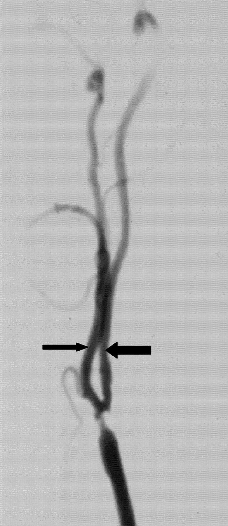
Methods: We re-reviewed 1216 patients with severe (> or =70%) stenosis on angiography in the North American Symptomatic Carotid Endarterectomy Trial and European Carotid Surgery Trial. One of 5 (n = 262) had 2 or more criteria for near occlusion: (1) delayed cranial arrival of ICA contrast compared with external carotid artery (ECA); (2) intracranial collaterals seen as cross-filling of contralateral vessels or ipsilateral contrast dilution; (3) obvious diameter reduction of ICA compared with opposite ICA; or (4) ICA diameter reduction compared with ipsilateral ECA.

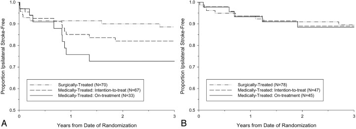
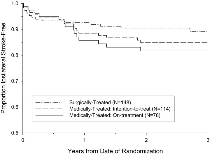
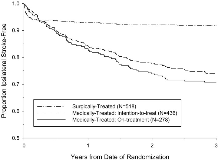
Results: Interrater agreement, sensitivity, and specificity were excellent (0.88, 90.6%, and 93.8%, respectively). By intention to treat, 3-year risks of ipsilateral stroke for medically treated patients with near occlusion was 15.1% versus 10.9% for surgically treated (absolute risk reduction [ARR] = 4.2%; P value = .33). Patients who continued to receive treatment in the medical arm for the trial's duration had a 3-year risk of 18.3% (ARR = 7.4%; P value = .13). Medically treated patients with severe stenosis but without near occlusion had a 3-year risk of 26.0% versus surgically treated of 8.2% (ARR = 17.8%; P value < .001).

Conclusion: It is crucial to identify near occlusions on vascular imaging. Although it still is reasonable to consider endarterectomy for these patients, the benefit is muted.


Figures

F<sc>ig</sc> 1.
Fig 1.
Near occlusion with collapsed lumen. Lateral common carotid angiogram shows the thin collapsed lumen (arrows) of the ICA above a prominent internal carotid stenosis at the carotid bulb (not shown).
F<sc>ig</sc> 2.
Fig 2.
Progression of stenosis to near occlusion. A, Anterioposterior angiogram shows ulcerated stenosis of the left carotid bulb. The normal ICA (larger arrow) beyond the stenosis is about twice the diameter of the ipsilateral distal ECA (smaller arrow). B, Anterioposterior angiogram about 1 year later shows progression of stenosis to near occlusion with the left ICA (larger arrow) reduced in diameter in comparison to previous angiogram and close to the diameter of the ECA (smaller arrow).
F<sc>ig</sc> 3.
Fig 3.
Progression of near occlusion to occlusion. A, Lateral carotid angiogram shows ulcerated stenosis causing near occlusion with ICA diameter beyond the stenosis (larger arrow), reduced in diameter to be smaller than the distal ECA diameter (smaller arrow). It should be about twice that diameter. B, Lateral carotid angiogram of the same carotid as A about 8 months later shows that the near occlusion has progressed to occlusion.
F<sc>ig</sc> 4.
Fig 4.
Collaterals (cross-filling and dilution) and late arrival of ICA to the head. A, Anterioposterior right carotid angiogram, head view, contralateral to left near occlusion shows exuberant cross-filling to left middle cerebral branches and even slightly down the supraclinoid carotid (arrow) to supply the anterior choroidal artery. B, Lateral left carotid angiogram, late arterial neck and head view, shows delay in ICA filling intracranially, and abrupt dilution contrast in the supraclinoid carotid (arrow) above the origin of a fetal-type posterior cerebral artery. This dilution indirectly demonstrates the excellent collateral already shown in A, not opacified here. It also shows the supraclinoid carotid site of the “watershed” between the right carotid source and the extremely severe left carotid stenosis.
F<sc>ig</sc> 5.
Fig 5.
Delayed ICA filling and intracranial dilution from collaterals. A, Lateral carotid angiogram (neck view) shows delayed ICA filling (larger arrow) beyond the nearly occluded bulb stenosis. B, There is dilution of contrast of middle cerebral branches (small arrows) intracranially, implying inflow of unopacified blood from circle of Willis collateral sources.
F<sc>ig</sc> 6.
Fig 6.
Decreased diameter of the ipsilateral ICA compared with the normal opposite carotid artery. A, Anterioposterior right carotid angiogram shows the diameter of the normal ICA (arrow). B, Anterioposterior left carotid angiogram done with the same magnification (same position of radiograph tube and image intensifier; same field of view) as the right in A showing decreased diameter of the left ICA (arrow).
F<sc>ig</sc> 7.
Fig 7.
Reduced ICA diameter compared with ECA. ICA diameter (larger arrow) is slightly less than distal ECA (small arrow), implying gross reduction of expected diameter. This implies that the ICA is much narrower than it should be.
F<sc>ig</sc> 8.
Fig 8.
Kaplan-Meier event-free survival from ipsilateral stroke for patients with near occlusion. A, The 3-year intention-to-treat risk of ipsilateral stroke in NASCET patients was 17.9% for medically treated and 11.4% for surgically treated. The 3-year risk of ipsilateral stroke for medically treated NASCET patients who continued to receive medical therapy throughout the duration of the trial (on-treatment) was 27.3%. B, The 3-year intention-to-treat risk of ipsilateral stroke in ECST patients was 11.1% for medically treated and 10.5% for surgically treated. The 3-year risk of ipsilateral stroke for medically treated ECST patients who continued to receive medical therapy throughout the duration of the trial (on-treatment) was 11.6%.
F<sc>ig</sc> 9.
Fig 9.
Kaplan-Meier event-free survival from ipsilateral stroke for patients with near occlusion, pooled data from the NASCET and ECST. The 3-year intention-to-treat risk of ipsilateral stroke in was 15.1% for medically treated and 10.9% for surgically treated. The 3-year risk of ipsilateral stroke for medically treated patients who continued to receive medical therapy throughout the duration of the trial (on-treatment) was 18.3%.
F<sc>ig</sc> 10.
Fig 10.
Kaplan-Meier event-free survival from ipsilateral stroke for patients with severe stenosis but without near occlusion, pooled data from the NASCET and ECST. The 3-year intention-to-treat risk of ipsilateral stroke in was 26.0% for medically treated and 8.2% for surgically treated. The 3-year risk of ipsilateral stroke for medically treated patients who continued to receive medical therapy throughout the duration of the trial (on-treatment) was 29.3%.

References

    1. Berman SS, Devine JJ, Erodes LS, Hunter GC. Distinguishing carotid artery pseudo-occlusion with color flow Doppler. Stroke 1995;26:434–438 - PubMed
    1. Dix JE, McNulty BJ, Kalimes DF. Frequency and significance of a small distal ICA in carotid stenosis. AJNR Am J Neuroradiol 1998;19:1215–1218 - PMC - PubMed
    1. Fox AJ. How to measure carotid stenosis. Radiology 1993;186:316–318 - PubMed
    1. Gabrielsen TO, Seeger JF, Knake JE, et al. The nearly occluded internal carotid artery: a diagnostic trap. Radiology 1981;138:611–618 - PubMed
    1. Henderson R, Eliasziw M, Fox AJ, et al. The importance of angiographically defined collateral circulation in patients with severe carotid stenosis. Stroke 2000;31:128–132 - PubMed

Publication types

LinkOut - more resources