. 2005 Sep 9;33(16):5106-11.
doi: 10.1093/nar/gki805. Print 2005.

Constraining ribosomal RNA conformational space

Affiliations

Constraining ribosomal RNA conformational space

Paola Favaretto et al. Nucleic Acids Res. .

Abstract

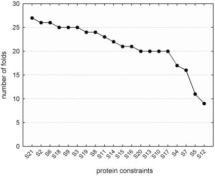
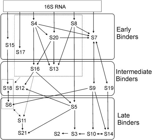
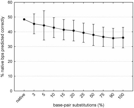
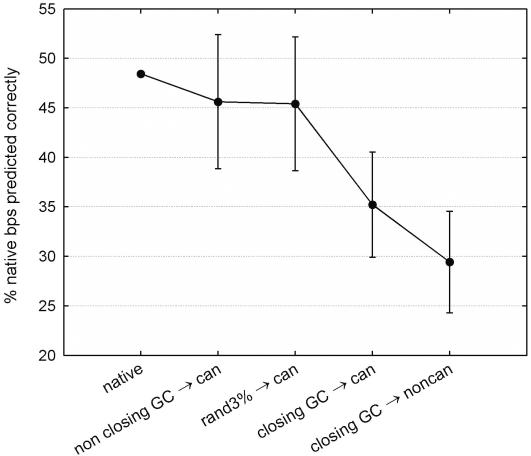
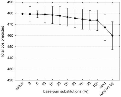
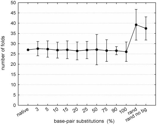
Despite the potential for many possible secondary-structure conformations, the native sequence of ribosomal RNA (rRNA) is able to find the correct and universally conserved core fold. This study reports a computational analysis investigating two mechanisms that appear to constrain rRNA secondary-structure conformational space: ribosomal proteins and rRNA sequence composition. The analysis was carried out by using rRNA-ribosomal protein interaction data for the Escherichia coli 16S rRNA and free energy minimization software for secondary-structure prediction. The results indicate that selection pressures on rRNA sequence composition and ribosomal protein-rRNA interaction play a key role in constraining the rRNA secondary structure to a single stable form.


Figures

Figure 1
Figure 1
Number of folds within 5% of the minimum free energy fold predicted when individual protein constraints were applied. In each case where a protein was reported to make contact with an rRNA residue that is part of a base pair in the native structure, the protein was assumed to force that particular residue to pair. If a protein makes contact with an rRNA residue that is part of a loop or bulge in the native structure, the protein was assumed to prevent that residue from pairing.
Figure 2
Figure 2
E.coli 16S ribosomal protein binding pathways determined from earlier in vitro studies (3,20). Arrows indicate ordered binding. Proteins are grouped together in terms of their temporal binding sequence as early, intermediate and late binders.
Figure 3
Figure 3
Average percentage of native base pairs predicted correctly for sequences with canonical base-pair substitutions. For each class of base-pair substitutions, the predicted base pairs that are present in the native structure are considered correct. The average is taken across the 100 samples of each class. A one-factor ANOVA test showed that the difference in the average percentage of base pairs correctly predicted is statistically significant (p < 0.05).
Figure 4
Figure 4
Average percentages of native base pairs predicted correctly for the following classes of sequences: ‘native’ sequence—16S rRNA sequence of E.coli; ‘non closing GC → can’—E.coli 16S rRNA sequences in which 3% of base pairs that are not G:C-tetraloop-closing base pairs are substituted by other canonical base pairs; ‘rand 3% → can’—E.coli 16S rRNA sequences in which ∼3% of the native base pairs are substituted by other canonical base pairs; ‘closing GC → can’—E.coli 16S rRNA sequences in which all the G:C-tetraloop-closing base pairs are substituted by other canonical base pairs (the total number of substitutions is equivalent to ∼3% of the total number of native base pairs); ‘closing GC → noncan’—E.coli 16S rRNA sequences in which all the G:C-tetraloop-closing base pairs are substituted by non-canonical base pairs (the total number of substitutions is equivalent to ∼3% of the native base pairs).
Figure 5
Figure 5
Average percentage of native base pairs predicted correctly for sequences with canonical base-pair substitutions. The native sequence is the 16S rRNA sequence of E.coli; ‘rand’ indicates random sequences of the same length and base distribution as the native sequence; ‘rand no bg’ indicates random sequences of the same length as the native sequence with a uniform base distribution.
Figure 6
Figure 6
Average number of secondary-structure folds predicted within 5% of the minimum free energy for sequences with canonical base-pair substitutions. Sequence categories are the same as in Figure 5.

References

    1. Cannone J.J., Subramanian S., Schnare M.N., Collett J.R., D'Souza L.M., Du Y., Feng B., Lin N., Madabusi L.V., Müller K.M., Pande N., Shang Z., Yu N., Gutell R.R. The Comparative RNA Web (CRW) site: an online database of comparative sequence and structure information for ribosomal, intron, and other RNAs. Bioinformatics. 2002;3:2. - PMC - PubMed
    1. Gutell R.R., Weiser B., Woese C.R., Noller H.F. Comparative anatomy of the 16-S-like ribosomal RNA. Prog. Nucleic Acid Res. Mol. Biol. 1985;32:155–216. - PubMed
    1. Brodersen D.E., Clemons W.E., Jr, Carter A.P., Wimberly B.T., Ramakrishnan V. Crystal structure of the 30S ribosomal subunit from Thermus thermophilus: structure of the proteins and their interactions with 16S RNA. J. Mol. Biol. 2002;316:725–768. - PubMed
    1. Klein D., Moore P., Steitz T. The roles of ribosomal proteins in the structure assembly, and evolution of the large ribosomal subunit. J. Mol. Biol. 2004;340:141–177. - PubMed
    1. Wimberly B.T., Brodersen D.E., Clemons W.M., Jr, Morgan-Warren R.J., Carter A.P., Vonrhein C., Hartsch T., Ramakrishnan V. Structure of the 30S ribosomal subunit. Nature. 2000;407:327–339. - PubMed

Publication types