

Structure of the nuclease domain of ribonuclease III from M. tuberculosis at 2.1 A
- PMID: 16155207
- PMCID: PMC2253305
- DOI: 10.1110/ps.051665905
Structure of the nuclease domain of ribonuclease III from M. tuberculosis at 2.1 A
Abstract
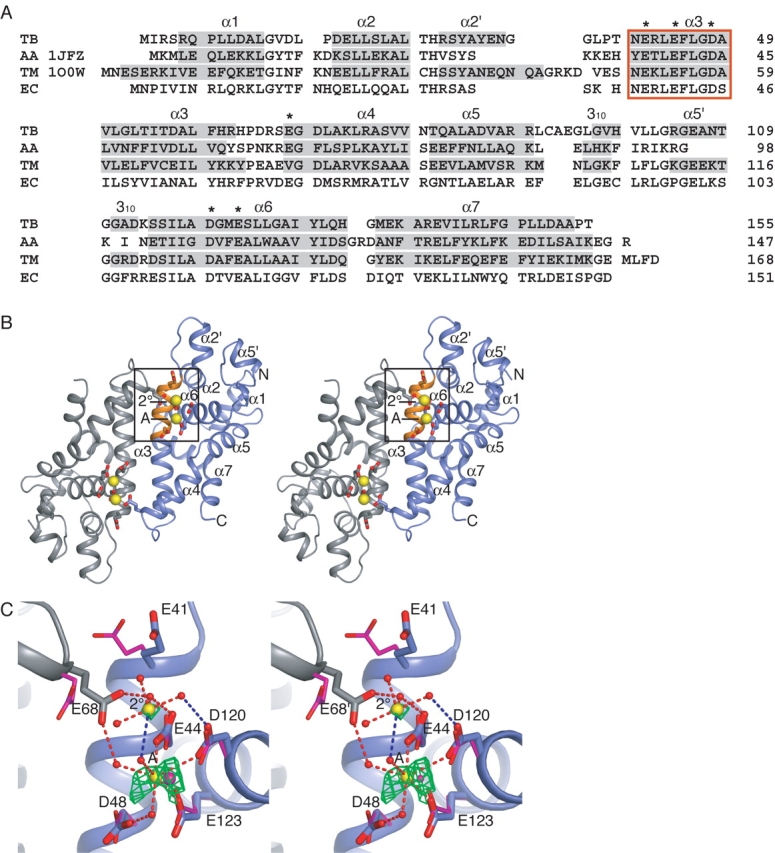
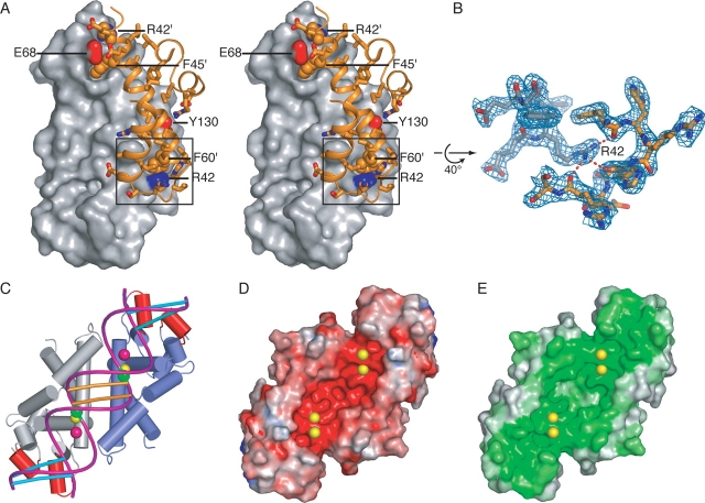
RNase III enzymes are a highly conserved family of proteins that specifically cleave double-stranded (ds)RNA. These proteins are involved in a diverse group of functions, including ribosomal RNA processing, mRNA maturation and decay, snRNA and snoRNA processing, and RNA interference. Here we report the crystal structure of the nuclease domain of RNase III from the pathogen Mycobacterium tuberculosis. Although globally similar to other RNase III folds, this structure has some features not observed in previously reported models. These include the presence of an additional metal ion near the catalytic site, as well as conserved secondary structural elements that are proposed to have functional roles in the recognition of dsRNAs.
Figures
References
-
- Blaszczyk, J., Tropea, J.E., Bubunenko, M., Routzahn, K.M., Waugh, D.S., Court, D.L., and Ji, X. 2001. Crystallographic and modeling studies of RNase III suggest a mechanism for double-stranded RNA cleavage. Structure (Camb) 9 1225–1236. - PubMed
-
- Blaszczyk, J., Gan, J., Tropea, J.E., Court, D.L., Waugh, D.S., and Ji, X. 2004. Noncatalytic assembly of ribonuclease III with double-stranded RNA. Structure (Camb) 12 457–466. - PubMed
-
- Brunger, A.T., Adams, P.D., Clore, G.M., DeLano, W.L., Gros, P., Grosse-Kunstleve, R.W., Jiang, J.S., Kuszewski, J., Nilges, M., Pannu, N.S., et al. 1998. Crystallography & NMR system: A new software suite for macromolecular structure determination. Acta Crystallogr. D Biol. Crystallogr. 54 (Pt. 5) 905–921. - PubMed
Publication types
MeSH terms
Substances
Associated data
- Actions
Grants and funding
LinkOut - more resources
Full Text Sources
