

Constitutive and accelerated shedding of murine syndecan-1 is mediated by cleavage of its core protein at a specific juxtamembrane site
- PMID: 16156648
- PMCID: PMC2546870
- DOI: 10.1021/bi050620i
Constitutive and accelerated shedding of murine syndecan-1 is mediated by cleavage of its core protein at a specific juxtamembrane site
Abstract
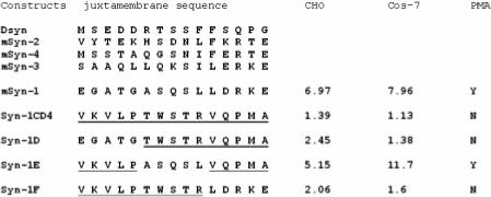
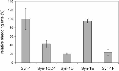
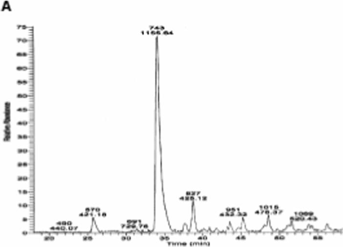
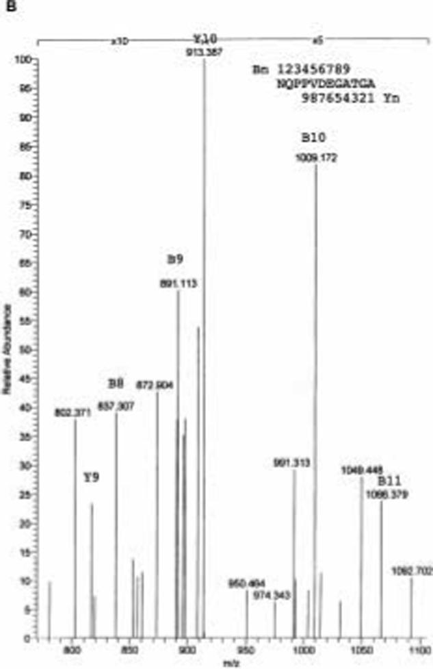
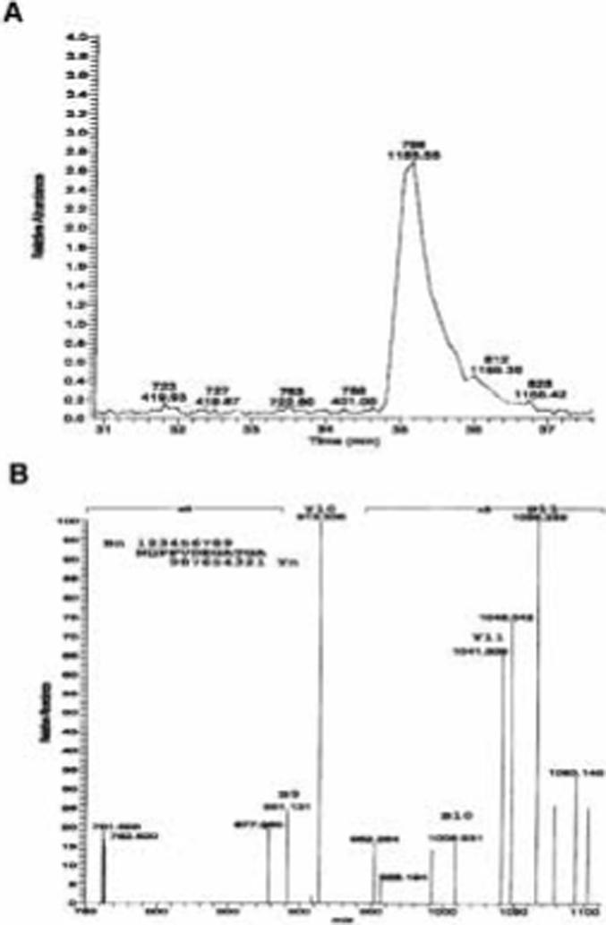
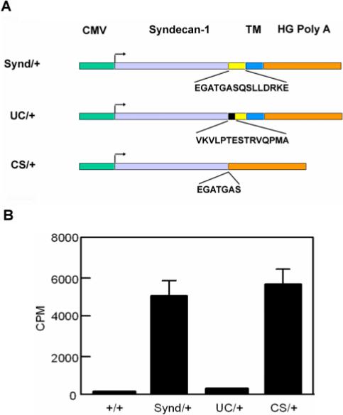
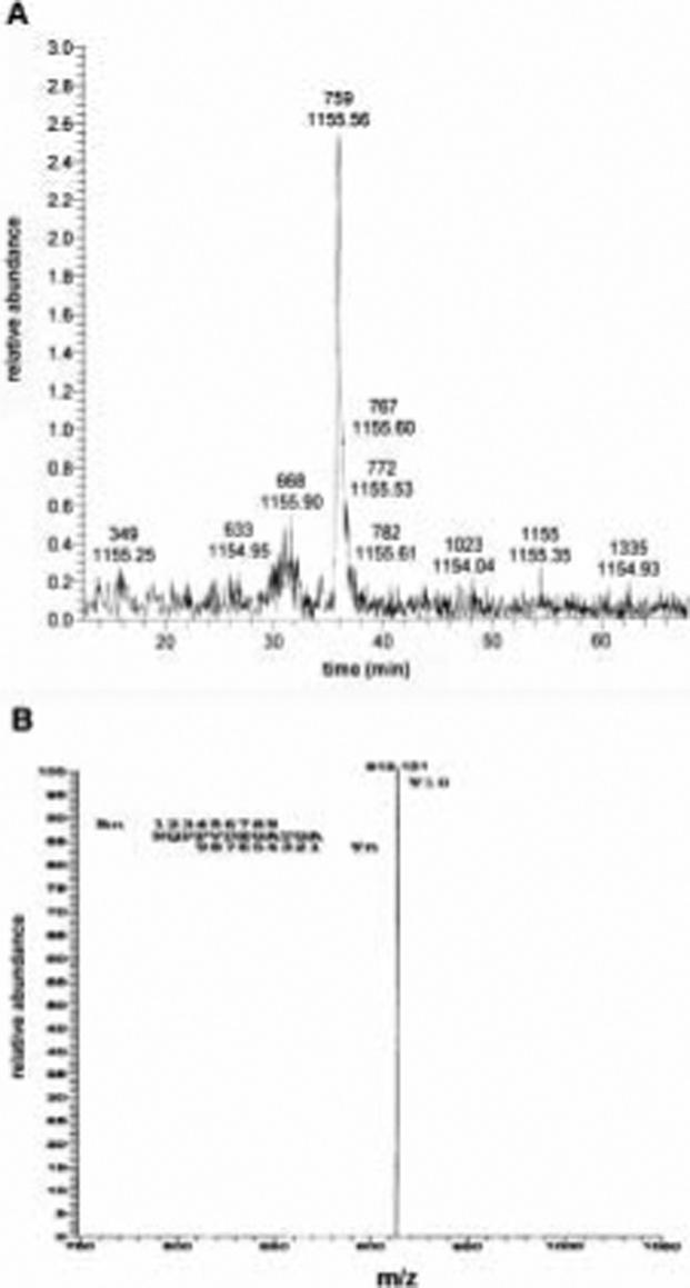
Syndecan-1 is a developmentally regulated cell surface heparan sulfate proteoglycan (HSPG). It functions as a coreceptor for a variety of soluble and insoluble ligands and is implicated in several biological processes, including differentiation, cell migration, morphogenesis, and recently feeding behavior. The extracellular domain of syndecan-1 is proteolytically cleaved at a juxtamembrane site by tissue inhibitor of metalloprotease-3 (TIMP-3)-sensitive metalloproteinases in response to a variety of physiological stimulators and stress in a process known as shedding. Shedding converts syndecan-1 from a membrane-bound coreceptor into a soluble effector capable of binding the same ligands. We found that replacing syndecan-1 juxtamembrane amino acid residues A243-S-Q-S-L247 with human CD4 amino acid residues can completely block PMA-induced syndecan-1 ectodomain shedding. Furthermore, using liquid chromatography-electrospray ionization mass spectrometry (LC-ESI-MS), we identified the proteolytic cleavage site of syndecan-1 as amino acids A243 and S244, generated by constitutive and PMA-induced shedding from murine NMuMG cells. Finally, we show that basal cleavage of syndecan-1 utilizes the same in vivo site as the in vitro site. Indeed, as predicted, transgenic mice expressing the syndecan-1/CD4 cDNA do not shed the syndecan-1 ectodomain in vivo. These results suggest that the same cleavage site is utilized for basal syndecan-1 ectodomain shedding both in vitro from NMuMG and CHO cells and in vivo.
Figures
Similar articles
-
Shedding of syndecan-1 and -4 ectodomains is regulated by multiple signaling pathways and mediated by a TIMP-3-sensitive metalloproteinase.J Cell Biol. 2000 Feb 21;148(4):811-24. doi: 10.1083/jcb.148.4.811. J Cell Biol. 2000. PMID: 10684261 Free PMC article.
-
Plasmin- and thrombin-accelerated shedding of syndecan-4 ectodomain generates cleavage sites at Lys(114)-Arg(115) and Lys(129)-Val(130) bonds.J Biol Chem. 2005 Oct 14;280(41):34441-6. doi: 10.1074/jbc.M501903200. Epub 2005 Aug 8. J Biol Chem. 2005. PMID: 16087677
-
Cleavage of syndecan-1 by membrane type matrix metalloproteinase-1 stimulates cell migration.J Biol Chem. 2003 Oct 17;278(42):40764-70. doi: 10.1074/jbc.M306736200. Epub 2003 Aug 6. J Biol Chem. 2003. PMID: 12904296
-
Syndecan, a developmentally regulated cell surface proteoglycan that binds extracellular matrix and growth factors.Philos Trans R Soc Lond B Biol Sci. 1990 Mar 12;327(1239):171-86. doi: 10.1098/rstb.1990.0052. Philos Trans R Soc Lond B Biol Sci. 1990. PMID: 1969657 Review.
-
Syndecans: multifunctional cell-surface co-receptors.Biochem J. 1997 Oct 1;327 ( Pt 1)(Pt 1):1-16. doi: 10.1042/bj3270001. Biochem J. 1997. PMID: 9355727 Free PMC article. Review.
Cited by
-
A transmembrane C-terminal fragment of syndecan-1 is generated by the metalloproteinase ADAM17 and promotes lung epithelial tumor cell migration and lung metastasis formation.Cell Mol Life Sci. 2015 Oct;72(19):3783-801. doi: 10.1007/s00018-015-1912-4. Epub 2015 Apr 26. Cell Mol Life Sci. 2015. PMID: 25912030 Free PMC article.
-
MMP7 shedding of syndecan-1 facilitates re-epithelialization by affecting alpha(2)beta(1) integrin activation.PLoS One. 2009 Aug 10;4(8):e6565. doi: 10.1371/journal.pone.0006565. PLoS One. 2009. PMID: 19668337 Free PMC article.
-
Extra-embryonic syndecan 2 regulates organ primordia migration and fibrillogenesis throughout the zebrafish embryo.Development. 2009 Sep;136(18):3143-52. doi: 10.1242/dev.031492. Development. 2009. PMID: 19700618 Free PMC article.
-
Heparan sulfate proteoglycans in cancer: Pathogenesis and therapeutic potential.Adv Cancer Res. 2023;157:251-291. doi: 10.1016/bs.acr.2022.08.001. Epub 2022 Aug 29. Adv Cancer Res. 2023. PMID: 36725112 Free PMC article.
-
Targeting syndecan-1: new opportunities in cancer therapy.Am J Physiol Cell Physiol. 2022 Jul 1;323(1):C29-C45. doi: 10.1152/ajpcell.00024.2022. Epub 2022 May 18. Am J Physiol Cell Physiol. 2022. PMID: 35584326 Free PMC article. Review.
References
-
- Bernfield M, Gotte M, Park PW, Reizes O, Fitzgerald ML, Lincecum J, Zako M. Functions of cell surface heparan sulfate proteoglycans. Annu Rev Biochem. 1999;68:729. - PubMed
-
- Gotte M, Joussen AM, Klein C, Andre P, Wagner DD, Hinkes MT, Kirchhof B, Adamis AP, Bernfield M. Role of syndecan-1 in leukocyte-endothelial interactions in the ocular vasculature. Invest Ophthalmol Vis Sci. 2002;43:1135. - PubMed
-
- Bernfield M, Kokenyesi R, Kato M, Hinkes MT, Spring J, Gallo RL, Lose EJ. Biology of the syndecans: a family of transmembrane heparan sulfate proteoglycans. Annu Rev Cell Biol. 1992;8:365. - PubMed
-
- Turner AJ, Hooper NM. Role for ADAM-family proteinases as membrane protein secretases. Biochem Soc Trans. 1999;27:255. - PubMed
-
- Werb Z, Yan Y. A cellular striptease act. Science. 1998;282:1279. - PubMed
Publication types
MeSH terms
Substances
Grants and funding
LinkOut - more resources
Full Text Sources
Other Literature Sources
Molecular Biology Databases
Research Materials
Miscellaneous
