. 2005 Sep 12:2:81.
doi: 10.1186/1743-422X-2-81.

Involvement of PKR and RNase L in translational control and induction of apoptosis after Hepatitis C polyprotein expression from a vaccinia virus recombinant

Affiliations

Involvement of PKR and RNase L in translational control and induction of apoptosis after Hepatitis C polyprotein expression from a vaccinia virus recombinant

Carmen E Gómez et al. Virol J. .

Abstract

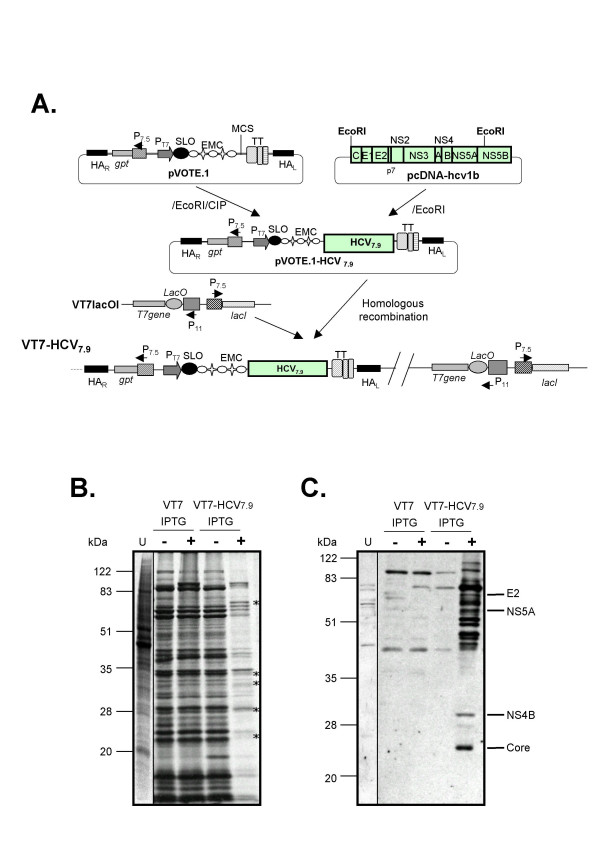
Background: Hepatitis C virus (HCV) infection is of growing concern in public health with around 350 million chronically infected individuals worldwide. Although the IFN-alpha/rivabirin is the only approved therapy with 10-30% clinical efficacy, the protective molecular mechanism involved during the treatment is still unknown. To analyze the effect of HCV polyprotein expression on the antiviral response of the host, we developed a novel vaccinia virus (VV)-based delivery system (VT7-HCV7.9) where structural and nonstructural (except part of NS5B) proteins of HCV ORF from genotype 1b are efficiently expressed and produced, and timely regulated in mammalian cell lines.

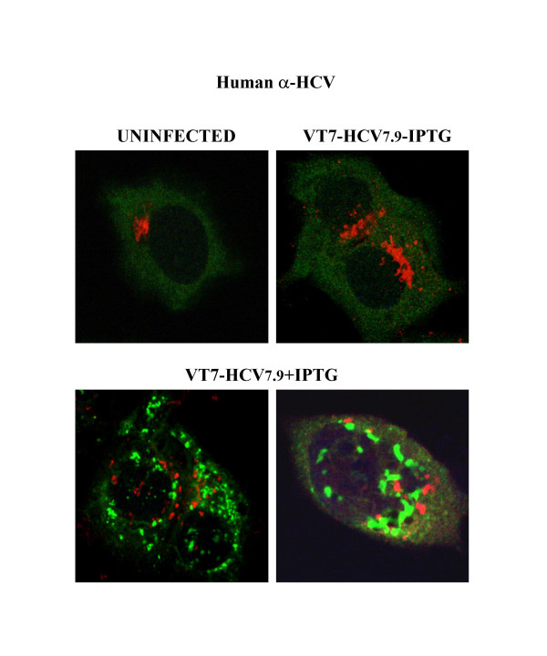
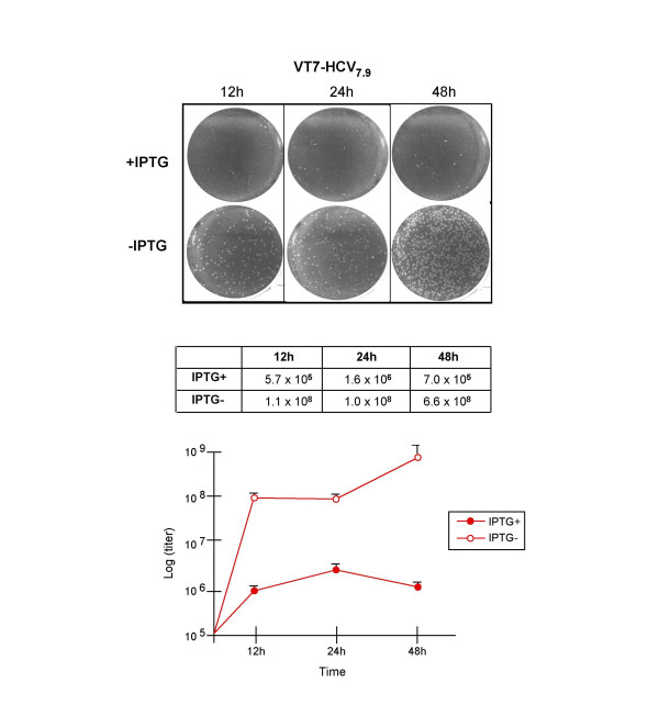
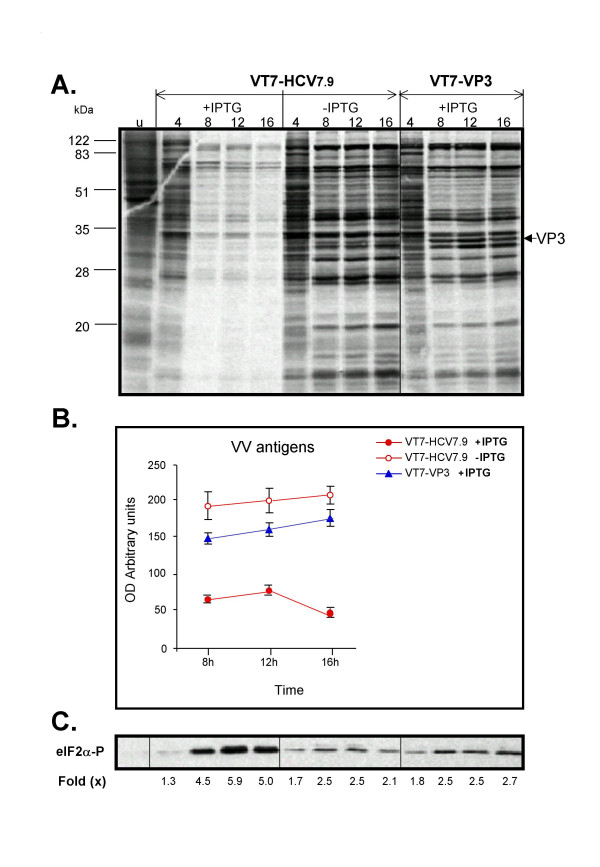
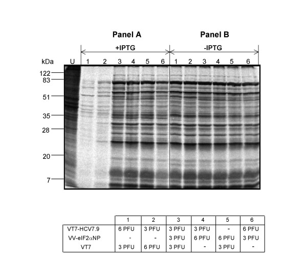
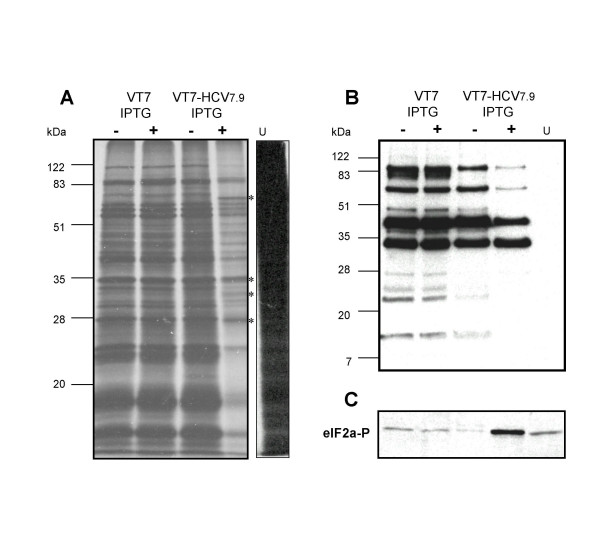
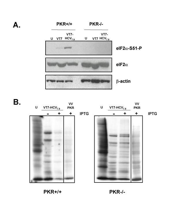
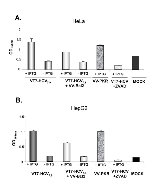
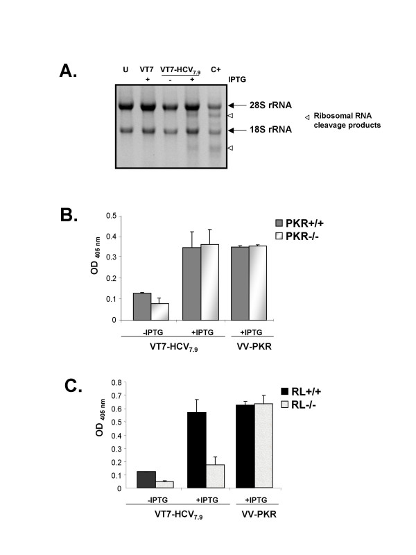
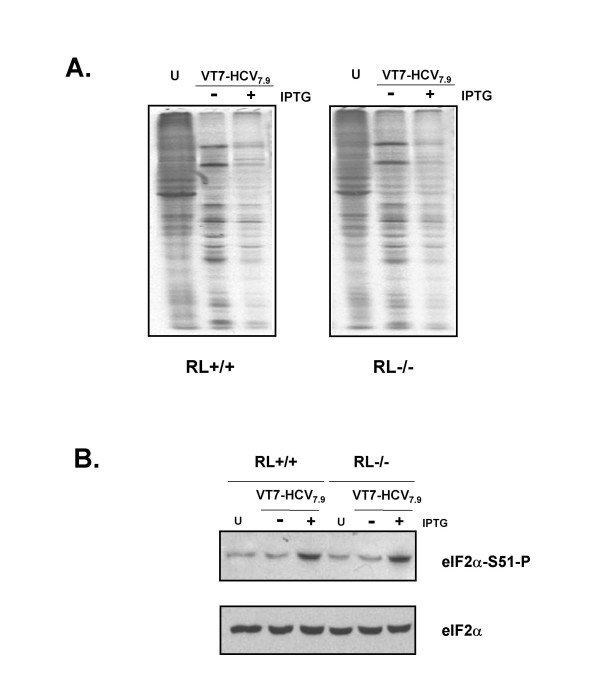
Results: Regulated transcript production and viral polypeptide processing was demonstrated in various cell lines infected with the recombinant VT7-HCV7.9, indicating that the cellular and viral proteolytic machineries are functional within these cells. The inducible expression of the HCV polyprotein by VV inhibits the synthesis of both host and viral proteins over the time and also induces apoptosis in HeLa and HepG2-infected cells. These effects occur accompanying with the phosphorylation of the translation initiation factor eIF-2alpha. In cells co-infected with VT7-HCV7.9 and a recombinant VV expressing the dominant negative eIF-2alpha-S51A mutant in the presence of the inductor isopropyl-thiogalactoside (IPTG), protein synthesis is rescued. The IFN-inducible protein kinase PKR is responsible for the translational block, as demonstrated with PKR-/- and PKR +/+ cell lines. However, apoptosis induced by VT7-HCV7.9 is mediated by the RNase L pathway, in a PKR-independent manner.

Conclusion: These findings demonstrate the antiviral relevance of the proteins induced by interferon, PKR and RNase L during expression from a VV recombinant of the HCV polyprotein in human cell lines. HCV polyprotein expression caused a severe cytopathological effect in human cells as a result of inhibition of protein synthesis and apoptosis induction, triggered by the activation of the IFN-induced enzymes PKR and RNase L systems. Thus, the virus-cell system described here highlights the relevance of the IFN system as a protective mechanism against HCV infection.


Figures

Figure 1
Figure 1
Construction and characterization of the recombinant VT7-HCV7.9 virus. A: Generation of recombinant VT7-HCV7.9. A 7.9 Kb DNA fragment containing the structural (C, E1, E2 and p7) and nonstructural (NS3, NS4A, NS4B, NS5A and the amino terminal region of NS5B) proteins of HCV from genotype 1b was cloned into a unique EcoRI restriction site of pVOTE.1 to make the plasmid transfer vector pVOTE.1-HCV7.9. BSC40 cells infected with the recombinant VT7lacOI (VT7), were transfected with the plasmid pVOTE.1-HCV7.9 as described in Materials and Methods to generate the recombinant VT7-HCV7.9. B: Expression of HCV inhibits protein synthesis in mammalian cells. Monolayers of BSC40 cells were infected at 5 PFU/cell with either the parental VT7 or the recombinant VT7-HCV7.9 viruses in the presence (+) or absence (-) of the inductor IPTG. Uninfected (U) and infected cells were metabolically labelled with 35S-Met-Cys Promix (100 μCi/mL) from 4 to 24 h.p.i. as described in Materials and Methods. Approximately 100 μg of total cell protein extracted from uninfected (U) and infected cells, was fractionated by SDS-PAGE followed by autoradiography. (*) represents new additional polypeptides corresponding to the HCV proteins. C: Inducible expression of HCV proteins by recombinant VT7-HCV7.9 virus. BSC40 cells were infected as described above. Total cell protein lysates from uninfected (U) and infected cells at 24 h.p.i. were analysed by Western blot using a human anti-HCV antibody from an infected patient. The protein band migration of Core, E2, NS4B and NS5A, as determined with specific antibodies, is indicated.
Figure 2
Figure 2
Cellular localization of HCV proteins by immunofluorescence microscopy. Subconfluent HeLa cells were infected at 5 PFU/cell with the recombinant VT7-HCV7.9 in the presence (+) or absence (-) of the inductor IPTG. At 16 h.p.i, cells were doubly labelled with polyclonal antibody anti-Gigantine to detect the Golgi complex (red) and a 1/200 dilution of serum from an HCV-infected patient (green) followed by the appropriate fluorescent secondary antibody and ToPro reagent.
Figure 3
Figure 3
Expression of HCV polyprotein inhibits the production of infectious VV. BSC40 cells were infected at 5 PFU/cell with the recombinant VT7-HCV7.9 in the presence or absence of IPTG. After the indicated times postinfection the cells were collected, centrifuged and resuspended in 300 μL of DMEM. After three freeze-thawing cycles, followed by sonication, the cell extracts were titrated in BSC40 cells. The experiment was performed two times in duplicate. Means and standard deviations are shown.
Figure 4
Figure 4
Time-course analysis of cellular and viral protein synthesis in cells expressing HCV polyprotein. A: BSC40 cells infected with the recombinant VT7-HCV7.9 virus in the presence (+) or absence (-) of IPTG were metabolically labelled with [35S] Met-Cys Promix (50 μCi/mL) at the indicated times (h.p.i) and analysed by SDS-PAGE (12%) and autoradiography. For comparative purposes, we included a similar inducible recombinant virus but expressing the IBDV mature structural capsid protein VP3 (VT7-VP3). B: Inhibition of VV proteins after expression of HCV. The levels of VV proteins were quantitated from autoradiograms using a BioRad GS700 image densitometer and computer software as suggested by the manufacturer. C: Immunoblot analysis of phospho-eIF-2α-S51 protein levels during the time-course of VT7-HCV7.9 infection. The number appearing in each lane represents the ratio of phospho-eIF-2α-S51 levels in infected cells compared to levels in uninfected cells.
Figure 5
Figure 5
Expression of the dominant negative eIF-2α-S51A mutant by VV-eIF2αNP rescues the translation inhibition induced by HCV polyprotein. BSC40 cells grown in 12-well plates were infected at a total of 9 PFU/cell with the viruses indicated in the presence or absence of IPTG (1.5 mM). At 18 h.p.i. the cells were metabolically labeled with [35S] Met-Cys Promix (50 μCi/mL) for 30 min. and analysed by SDS-PAGE (12%) and autoradiography.
Figure 6
Figure 6
Expression of HCV polyprotein from VV inhibits cellular and viral protein synthesis in the hepatic cell line HepG2. A: Monolayers of HepG2 cells were infected (5 PFU/cell) with either VT7 or VT7-HCV7.9 recombinant viruses, in the presence (+) or absence (-) of the inductor IPTG. Uninfected (U) and infected cells were metabolically labelled with [35S] Met-Cys Promix (100 μCi/mL) from 4 to 24 h.p.i and treated as described under Materials and Methods. Approximately 100 μg of total cell protein extracted from uninfected and infected cells was fractionated by SDS-PAGE followed by autoradiography. (*) represents new additional polypeptides corresponding to the HCV proteins. B: Immunoblot analysis of total cell protein lysates prepared from uninfected and infected cells at 24 h.p.i. The blot was probed with a rabbit polyclonal anti-serum raised against live VV. C: The blot was stripped and probed again with a polyclonal antibody that recognized phospho-eIF-2α-S51 protein.
Figure 7
Figure 7
PKR mediates phosphorylation of eIF-2α and inhibition of translation caused by the expression of HCV polyprotein. A: Immunoblot analysis of total cell protein lysates prepared from PKR knockout (PKR-/-) and PKR WT (PKR+/+) cells infected with the parental (VT7) or the recombinant VT7-HCV7.9 viruses in the presence (+) of IPTG for 24 h. The blot was first probed with a polyclonal antibody that recognized phospho-eIF-2α-S51 protein, stripped twice, and reprobed with a polyclonal antibody that recognizes total eIF-2α protein and a monoclonal antibody against β-actin. B: Wild type and PKR-/- cell lines infected with VT7-HCV7.9 in the presence (+) or absence (-) of IPTG were metabolically labelled with 35S-Met-Cys Promix (50 μCi/mL) at 16 h.p.i, fractionated by SDS-PAGE and analysed by autoradiography. The recombinant VV-PKR virus was used as a control. U: uninfected cells.
Figure 8
Figure 8
Expression of HCV polyprotein from VV induces apoptosis in HeLa and HepG2 cells that is caspase-dependent. A: HeLa cells were infected at 5 PFU/cell with the recombinant VT7-HCV7.9 individually or in combination (2.5 PFU of each virus/cell) with the recombinant VV-Bcl2 (inducibly expressing the anti-apoptotic Bcl-2 polypeptide) or with a general caspase inhibitor, Z-VAD-FMK (Calbiochem) at 50 μM, in the presence (+) or absence (-) of IPTG. The apoptotic levels were determined at 24 h.p.i by ELISA. B: HepG2 cells were infected at 10 PFU/cell with the recombinant VT7-HCV7.9 individually or in combination (5 PFU of each virus/cell) with the recombinant VV-Bcl2 or with a general caspase inhibitor, Z-VAD-FMK (Calbiochem) at 50 μM, in the presence (+) or absence (-) of IPTG. The apoptotic levels were determined at 48 h.p.i by ELISA. VV-PKR infected cells in the presence (+) of IPTG were used as positive controls.
Figure 9
Figure 9
Expression of HCV polyprotein from VV induces ribosomal RNA degradation mediated by RNaseL and triggers apoptosis through RNase L independently of PKR. A: Monolayers of HeLa cells were either uninfected (U), single-infected with VT7 (5 PFU/cell), single-infected with VT7-HCV7.9 (5 PFU/cell) in the presence (+) or absence (-) of IPTG, or triple-infected with VV-RL + VT7 + VV-25AS (2 PFU of each virus/cell) (C+). Infections proceeded for 24 hours. 2 μg of total RNA was fractionated in 1% agarose-formaldehyde gel and stained with ethidium bromide. Abundant ribosomal RNAs 28S and 18S are indicated. B and C: PKR knockout (PKR-/-) and PKR WT cells (PKR+/+) (panel B), as well as RNase L knockout (RL-/-) and RNase L WT cells (RL+/+) (panel C), were infected at 5 PFU/cell with the recombinant VT7-HCV7.9 virus, in the presence (+) or absence (-) of the inductor IPTG. The apoptotic levels in cell extracts were determined at 24 h.p.i. by ELISA. The recombinant VV-PKR virus was used as a control. U: Uninfected cells.
Figure 10
Figure 10
Expression of HCV polyprotein from VV inhibits cellular and viral protein synthesis in RL+/+ and in RL-/- infected cells. A: RL+/+ and RL-/- cells infected with VT7-HCV7.9 in the presence (+) or absence (-) of IPTG were metabolically labelled with 35S-Met-Cys Promix (50 μCi/mL) at 8 h.p.i, fractionated by SDS-PAGE and analysed by autoradiography. U: uninfected cells. B: Immunoblot analysis of total cell protein lysates prepared from RL+/+ and RL-/- cells infected with VT7-HCV7.9 in the presence (+) or absence (-) of IPTG for 8 h. The blot was first probed with a polyclonal antibody that recognized phospho-eIF-2α-S51 protein, stripped and reprobed with a polyclonal antibody that recognizes total eIF-2α protein.

Similar articles

Cited by

References

    1. Choo QL, Kuo G, Weiner AJ, Overby LR, Bradley DW, Houghton M. Isolation of a cDNA clone derived from a blood-borne non-A, non-B viral hepatitis genome. Science. 1989;244:359–362. - PubMed
    1. Kuo G, Choo QL, Alter HJ, Gitnick GL, Redeker AG, Purcell RH, Miyamura T, Dienstag JL, Alter MJ, Stevens CE, et al. An assay for circulating antibodies to a major etiologic virus of human non-A, non-B hepatitis. Science. 1989;244:362–364. - PubMed
    1. Robertson B, Myers G, Howard C, Brettin T, Bukh J, Gaschen B, Gojobori T, Maertens G, Mizokami M, Nainan O, Netesov S, Nishioka K, Shin i T, Simmonds P, Smith D, Stuyver L, Weiner A. Classification, nomenclature, and database development for hepatitis C virus (HCV) and related viruses: proposals for standardization. International Committee on Virus Taxonomy. Arch Virol. 1998;143:2493–2503. doi: 10.1007/s007050050479. - DOI - PubMed
    1. Bartenschlager R. The NS3/4A proteinase of the hepatitis C virus: unravelling structure and function of an unusual enzyme and a prime target for antiviral therapy. J Viral Hepat. 1999;6:165–181. doi: 10.1046/j.1365-2893.1999.00152.x. - DOI - PubMed
    1. Reed KE, Rice CM. Overview of hepatitis C virus genome structure, polyprotein processing, and protein properties. Curr Top Microbiol Immunol. 2000;242:55–84. - PubMed

Publication types

LinkOut - more resources