

The interaction of the SARS coronavirus non-structural protein 10 with the cellular oxido-reductase system causes an extensive cytopathic effect
- PMID: 16157265
- PMCID: PMC7108382
- DOI: 10.1016/j.jcv.2004.12.019
The interaction of the SARS coronavirus non-structural protein 10 with the cellular oxido-reductase system causes an extensive cytopathic effect
Abstract
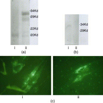
The pathological mechanism of SARS-CoV infection was investigated. The gene for the SARS-CoV non-structural protein 10, which is located in the open reading frame of pp1a/pp1ab gene, was synthesized and used to screen for the specific cellular gene coding for the protein interacting with this nsp10 protein in a human embryo lung cDNA library using a yeast trap method. The results indicated that apart from the two subunits of cellular RNA polymerase complex, BTF3 and ATF5, this nsp10 protein was also able to interact specifically with the NADH 4L subunit and cytochrome oxidase II. Further study revealed that the activity of the NADH-cytochrome was altered and the inner mitochondrial membrane was depolarized in the transfected human embryo lung fibroblast by the nsp10 protein gene. The cytopathic effect of the Coronavirus 229E strain appeared more extensive in these cells than in the control cells.
Figures


Similar articles
-
Biochemical and structural insights into the mechanisms of SARS coronavirus RNA ribose 2'-O-methylation by nsp16/nsp10 protein complex.PLoS Pathog. 2011 Oct;7(10):e1002294. doi: 10.1371/journal.ppat.1002294. Epub 2011 Oct 13. PLoS Pathog. 2011. PMID: 22022266 Free PMC article.
-
[Screening and cloning of hepatitis C virus non-structural protein 4B interacting protein gene in hepatocytes].Zhonghua Shi Yan He Lin Chuang Bing Du Xue Za Zhi. 2005 Sep;19(3):248-51. Zhonghua Shi Yan He Lin Chuang Bing Du Xue Za Zhi. 2005. PMID: 16261208 Chinese.
-
The SARS-Coronavirus PLnc domain of nsp3 as a replication/transcription scaffolding protein.Virus Res. 2008 May;133(2):136-48. doi: 10.1016/j.virusres.2007.11.017. Epub 2008 Feb 5. Virus Res. 2008. PMID: 18255185 Free PMC article.
-
Design and Evaluation of Anti-SARS-Coronavirus Agents Based on Molecular Interactions with the Viral Protease.Molecules. 2020 Aug 27;25(17):3920. doi: 10.3390/molecules25173920. Molecules. 2020. PMID: 32867349 Free PMC article. Review.
-
Role of Structural and Non-Structural Proteins and Therapeutic Targets of SARS-CoV-2 for COVID-19.Cells. 2021 Apr 6;10(4):821. doi: 10.3390/cells10040821. Cells. 2021. PMID: 33917481 Free PMC article. Review.
Cited by
-
Severe acute respiratory syndrome coronavirus as an agent of emerging and reemerging infection.Clin Microbiol Rev. 2007 Oct;20(4):660-94. doi: 10.1128/CMR.00023-07. Clin Microbiol Rev. 2007. PMID: 17934078 Free PMC article. Review.
-
Myocardial Damage by SARS-CoV-2: Emerging Mechanisms and Therapies.Viruses. 2021 Sep 21;13(9):1880. doi: 10.3390/v13091880. Viruses. 2021. PMID: 34578462 Free PMC article. Review.
-
Upregulation of mitochondrial gene expression in PBMC from convalescent SARS patients.J Clin Immunol. 2006 Nov;26(6):546-54. doi: 10.1007/s10875-006-9046-y. Epub 2006 Oct 6. J Clin Immunol. 2006. PMID: 17024565 Free PMC article.
-
Crystal structure of nonstructural protein 10 from the severe acute respiratory syndrome coronavirus reveals a novel fold with two zinc-binding motifs.J Virol. 2006 Aug;80(16):7894-901. doi: 10.1128/JVI.00467-06. J Virol. 2006. PMID: 16873246 Free PMC article.
-
How the Competition for Cysteine May Promote Infection of SARS-CoV-2 by Triggering Oxidative Stress.Antioxidants (Basel). 2023 Feb 14;12(2):483. doi: 10.3390/antiox12020483. Antioxidants (Basel). 2023. PMID: 36830041 Free PMC article.
References
-
- Anand K., Ziebuhr J., Wadhwani P., Mesters J.R., Hilgenfeld R. Coronavirus main proteinase (3clpro) structure: basis for design of anti-SARS drugs. Science. 2003;300:1763–1767. - PubMed
-
- Booth C.M., Matukas L.M., Tomlinson G.A., Rachlis A.R., Rose D.B., Dwosh H.A. Clinical features and short-term outcomes of 144 patients with SARS in the Greater Toronto area. JAMA. 2003;289:2801–2809. - PubMed
-
- Fields S., Sternglanz R. The two-hybrid system: an assay for protein–protein interactions. Trends Genet. 1994;10:286–292. - PubMed
Publication types
MeSH terms
Substances
Grants and funding
LinkOut - more resources
Full Text Sources
Other Literature Sources
Molecular Biology Databases
Miscellaneous
