

Localization of a disease-associated mutation site in the three-dimensional structure of the cardiac muscle ryanodine receptor
- PMID: 16157601
- PMCID: PMC1470666
- DOI: 10.1074/jbc.M505714200
Localization of a disease-associated mutation site in the three-dimensional structure of the cardiac muscle ryanodine receptor
Abstract
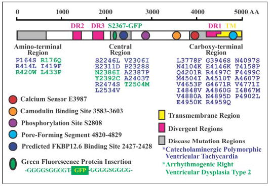
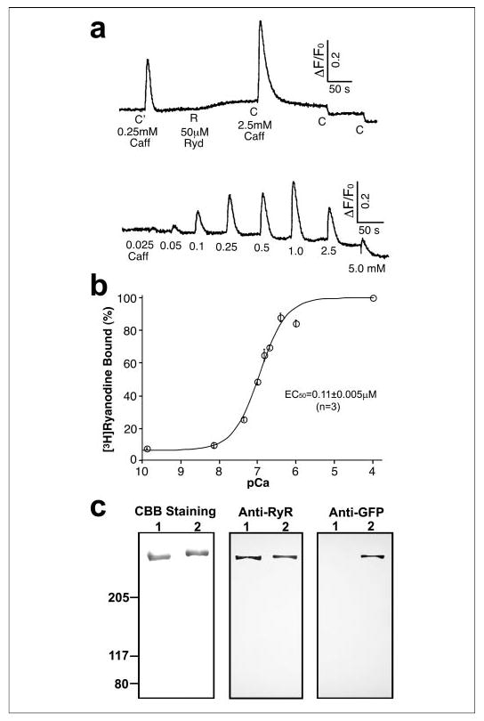
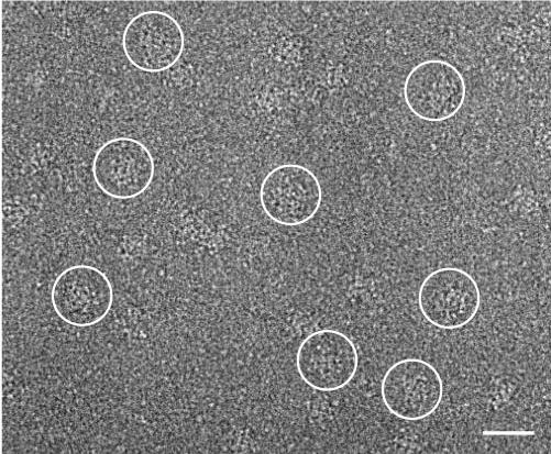
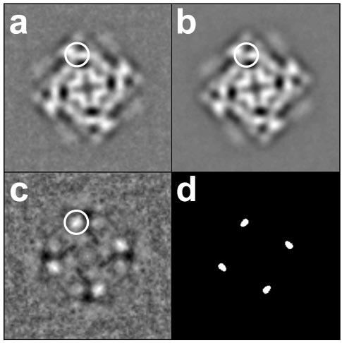
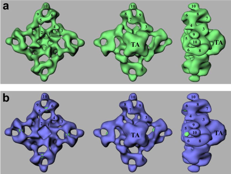
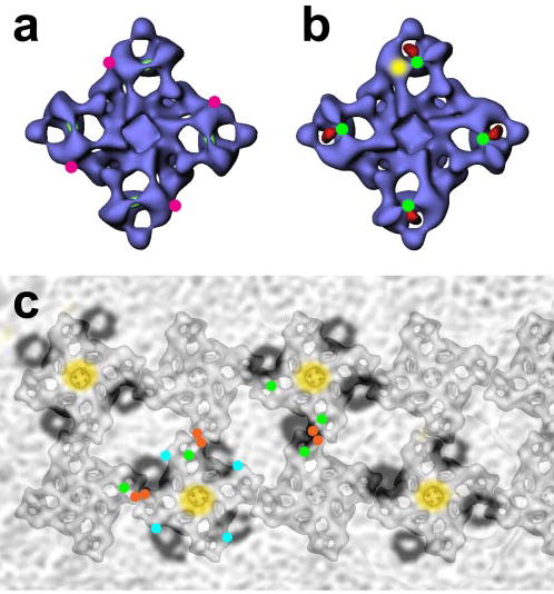
The cardiac muscle ryanodine receptor (RyR2) functions as a calcium release channel in the heart. Up to 40 mutations in RyR2 have been linked to genetic forms of sudden cardiac death. These mutations are largely clustered in three regions of the sequence of the polypeptide: one near the N terminus, one in the central region, and the third in the C-terminal region. The central region includes 11 mutations, and an isoleucine-proline motif (positions 2427 and 2428) in the same region is predicted to contribute to the binding of FKBP12.6 protein. We have mapped the central mutation site in the three-dimensional structure of RyR2 by green fluorescent protein insertion, cryoelectron microscopy, and single-particle image processing. The central mutation site was mapped to a "bridge" of density that connects cytoplasmic domains 5 and 6, which have been suggested to undergo conformational changes during channel gating. Moreover, the location of this central mutation site is not close to that of the FKBP12.6-binding site mapped previously by cryoelectron microscopy.
Figures
Similar articles
-
Localization of PKA phosphorylation site, Ser(2030), in the three-dimensional structure of cardiac ryanodine receptor.Biochem J. 2008 Mar 1;410(2):261-70. doi: 10.1042/BJ20071257. Biochem J. 2008. PMID: 17967164 Free PMC article.
-
Localization of an NH(2)-terminal disease-causing mutation hot spot to the "clamp" region in the three-dimensional structure of the cardiac ryanodine receptor.J Biol Chem. 2007 Jun 15;282(24):17785-93. doi: 10.1074/jbc.M700660200. Epub 2007 Apr 23. J Biol Chem. 2007. PMID: 17452324 Free PMC article.
-
Three-dimensional localization of divergent region 3 of the ryanodine receptor to the clamp-shaped structures adjacent to the FKBP binding sites.J Biol Chem. 2003 Apr 18;278(16):14211-8. doi: 10.1074/jbc.M213164200. Epub 2003 Feb 7. J Biol Chem. 2003. PMID: 12576471
-
Involvement of the cardiac ryanodine receptor/calcium release channel in catecholaminergic polymorphic ventricular tachycardia.J Cell Physiol. 2002 Jan;190(1):1-6. doi: 10.1002/jcp.10031. J Cell Physiol. 2002. PMID: 11807805 Review.
-
Molecular regulation of cardiac ryanodine receptor ion channel.Cell Calcium. 2004 Jun;35(6):621-8. doi: 10.1016/j.ceca.2004.01.015. Cell Calcium. 2004. PMID: 15110152 Review.
Cited by
-
Single-particle cryo-EM of the ryanodine receptor channel in an aqueous environment.Eur J Transl Myol. 2015;25(1):4803. doi: 10.4081/ejtm.2015.4803. Eur J Transl Myol. 2015. PMID: 25844145 Free PMC article.
-
Ryanodine receptors: structure, expression, molecular details, and function in calcium release.Cold Spring Harb Perspect Biol. 2010 Nov;2(11):a003996. doi: 10.1101/cshperspect.a003996. Epub 2010 Oct 20. Cold Spring Harb Perspect Biol. 2010. PMID: 20961976 Free PMC article. Review.
-
ARVC-related mutations in divergent region 3 alter functional properties of the cardiac ryanodine receptor.Biophys J. 2008 Jun;94(12):4668-77. doi: 10.1529/biophysj.107.122382. Epub 2008 Mar 7. Biophys J. 2008. PMID: 18326664 Free PMC article.
-
Localization of PKA phosphorylation site, Ser(2030), in the three-dimensional structure of cardiac ryanodine receptor.Biochem J. 2008 Mar 1;410(2):261-70. doi: 10.1042/BJ20071257. Biochem J. 2008. PMID: 17967164 Free PMC article.
-
Catecholaminergic polymorphic ventricular tachycardia is caused by mutation-linked defective conformational regulation of the ryanodine receptor.Circ Res. 2010 Apr 30;106(8):1413-24. doi: 10.1161/CIRCRESAHA.109.209312. Epub 2010 Mar 11. Circ Res. 2010. PMID: 20224043 Free PMC article.
References
-
- American Heart Association (2005) Heart Disease and Stroke Statistics–2005 Update, American Heart Association, Dallas, TX
-
- Chugh SS, Senashova O, Watts A, Tran PT, Zhou Z, Gong Q, Titus JL, Hayflick SJ. J Am Coll Cardiol. 2004;43:1625–1629. - PubMed
-
- Priori SG, Napolitano C, Tiso N, Memmi M, Vignati G, Bloise R, Sorrentino V, Danieli GA. Circulation. 2001;103:196–200. - PubMed
-
- Laitinen PJ, Brown KM, Piippo K, Swan H, Devaney JM, Brahmbhatt B, Donarum EA, Marino M, Tiso N, Viitasalo M, Toivonen L, Stephan DA, Kontula K. Circulation. 2001;103:485–490. - PubMed
-
- Tiso N, Stephan DA, Nava A, Bagattin A, Devaney JM, Stanchi F, Larderet G, Brahmbhatt B, Brown K, Bauce B, Muriago M, Basso C, Thiene G, Danieli GA, Rampazzo A. Hum Mol Genet. 2001;10:189–194. - PubMed
Publication types
MeSH terms
Substances
Grants and funding
LinkOut - more resources
Full Text Sources
