. 2005 Sep 12;170(6):993-1004.
doi: 10.1083/jcb.200507082.

Stable interaction between alpha5beta1 integrin and Tie2 tyrosine kinase receptor regulates endothelial cell response to Ang-1

Affiliations

Stable interaction between alpha5beta1 integrin and Tie2 tyrosine kinase receptor regulates endothelial cell response to Ang-1

Ilaria Cascone et al. J Cell Biol. .

Abstract

During angiogenic remodeling, Ang-1, the ligand of Tie2 tyrosine kinase, is involved in vessel sprouting and stabilization through unclear effects on nascent capillaries and mural cells. In our study, we hypothesized that the Ang-1/Tie2 system could cross-talk with integrins, and be influenced by the dynamic interactions between extracellular matrix and endothelial cells (ECs). Here, we show that alpha5beta1 specifically sensitizes and modulates Tie2 receptor activation and signaling, allowing EC survival at low concentrations of Ang-1 and inducing persistent EC motility. Tie2 and alpha5beta1 interact constitutively; alpha5beta1 binding to fibronectin increases this association, whereas Ang-1 stimulation recruits p85 and FAK to this complex. Furthermore, we demonstrate that Ang-1 is able to mediate selectively alpha5beta1 outside-in FAK phosphorylation. Thus, Ang-1 triggers signaling pathways through Tie2 and alpha5beta1 receptors that could cross-talk when Tie2/alpha5beta1 interaction occurs in ECs plated on fibronectin. By using blocking antibodies, we consistently found that alpha5beta1, but not alphavbeta3 activation, is essential to Ang-1-dependent angiogenesis in vivo.


Figures

Figure 1.
Figure 1.
α5β1 engagement influences Tie2 phosphorylation and signaling. ECs cultured on native ECM (a) and (g), or on 5 μg/ml fibronectin (FN), 5 μg/ml collagen I (COLL), or 20 μg/ml fibrinogen (FB) (c, e, and g) were stimulated with Ang-1 for 5 min (a, c, and e) or VEGF-A165 (g) at the indicated concentrations, lysed, immunoprecipitated (IP) with either anti-Tie2 (a and c), anti–VEGFR-2 (g), or anti-p85 (c) and blotted as indicated. Panels b, d, and f show the densitometric analysis of a, c, and e, respectively, and indicate the fold increase of Tie2 or p85 tyrosine phosphorylation normalized to the total protein amount. Values shown are means ± SD of five independent experiments. In b, statistical significance (*, P < 0.01) is shown for increasing Ang-1 concentrations compared with 20 ng/ml Ang-1. In d and f, statistical significance (*, P < 0.01) is shown for Ang-1–stimulated ECs for the indicated substrate.
Figure 2.
Figure 2.
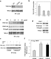
Tie2 and α5β1 interact. ECs cultured on native ECM were lysed and immunoprecipitated by anti-α5β1 (a), anti-α2β1 (b), or anti-αvβ3 (c) Abs or control immunoglobulins, and blotted with anti-Tie2 (a–c) or anti-β1 or anti-VEGFR-2 (a) or anti-α2 (b) or anti-β3 (c) Abs. Anti-α5β1 immunoprecipitates were re-immunoprecipitated (Re-IP) by anti-Tie2 Ab or control immunoglobulins and blotted with anti-Tie2 (d). CHO B2 and B2a27 cells expressing Tie2 were lysed, immunoprecipitated with anti-β1 Ab, and blotted with anti-Tie2 Ab (e), or biotinylated before lysis, Tie2 or control immunoglobulins re-immunoprecipitated and revealed by peroxidase-conjugated streptavidin (f). CHO B2 and B2a27 cells expressing Tie2 or VEGFR-2 adhered on poly-lysine (CHO B2) or on fibronectin (CHO B2a27) were stimulated with Ang-1 (50 ng/ml) or VEGF-A165 at the indicated time points; lysed; and immunoprecipitated with anti-Tie2 or anti-VEGFR-2 Abs and blotted with anti-phosphotyrosine (g and i), anti-Tie2 (g) or anti-VEGFR-2 Abs (i). h and l show the densitometric analysis where the Tie2 or VEGFR-2 tyrosine phosphorylation fold increase has been calculated as described in this legend. Values shown are means ± SD of five independent experiments. Statistical significance (*, P < 0.01) is shown for Ang-1– or VEGF-A165–stimulated cells compared with unstimulated cells.
Figure 3.
Figure 3.
Tie2/α5β1 complex increases upon α5β1 engagement and transduces upon Ang-1 stimulation. ECs plated on 5 μg/ml fibronectin, 5 μg/ml collagen I, and 20 μg/ml fibrinogen (a) or on increasing concentrations of fibronectin (1–10 μg/ml; c), or on 5 μg/ml fibronectin (d) were lysed, immunoprecipitated by anti-α5β1 Ab. (d) ECs were stimulated by Ang-1 (50 ng/ml) for 5 min. Blots were probed as indicated. CHO B2 and B2a27 were plated on poly-lysine or fibronectin (e), stimulated or not with Ang-1 (50 ng/ml) for 5 min, lysed, immunoprecipitated with anti-FAK, and blotted with antiphosphotyrosine or anti-FAK. Densitometric analysis shows the relative amount of Tie2 in α5β1 immunoprecipitates expressed in arbitrary unit (b) and the fold increase of FAK phosphorylation (f). Values shown are means ± SD of five independent experiments. Statistical significance (*, P < 0.01) is shown for ECs plated on fibronectin compared with the other conditions, and for Ang-1– or fibronectin-stimulated CHOB2a27 compared with CHO B2.
Figure 4.
Figure 4.
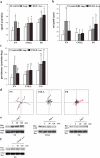
Ang-1 increases the speed and the persistence of EC plated on fibronectin. Starved ECs were plated on 5 μg/ml fibronectin (FN), 5 μg/ml collagen I (COLL), and 20 μg/ml fibrinogen (FG) for 1 h and stimulated or not with 50 ng/ml of Ang-1 or 10 ng/ml of VEGF-A165. Time-lapse videomicroscopy was performed and recorded images every 5 min for 4 h. 50 cells from five different videos were examined for speed (a), net path length covered (b), and persistence (c; deg, direction). (d) ECs paths were tracked over 4 h and replotted such that all paths start from origin. Values shown are means ± SD of 50 cells from five independent experiments. Statistical significance is shown (*, P < 0.01 and §, P < 0.05) for stimulated cells compared with unstimulated cells; ••, P < 0.01 for Ang-1–stimulated cells compared with VEGF-A165–stimulated cells. ECs plated as in (a) were stimulated with 50 ng/ml of Ang-1 (e) or 10 ng/ml of VEGF-A165 (f) for the indicated times, and cell lysates were incubated with GST-PBD to pull-down Rac1-GTP. Proteins separated by SDS-PAGE were probed with anti-Rac1 Ab. This experiment is representative of five experiments performed with similar results.
Figure 5.
Figure 5.
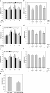
Ang-1 induces EC adhesion through specific integrin function. Panels a, c and e show the fold increase of EC adhesion induced by Ang-1 (20, 50, 100 ng/ml) versus unstimulated ECs to different concentrations of fibronectin (a), collagen I (c), or fibrinogen (e). Values shown are means ± SD of five independent experiments, each in triplicate. Statistical significance (*, P < 0.01, §, P < 0.05) is shown for increasing Ang-1 concentrations compared with 20 ng/ml Ang-1. b, d, and f show the fold increase of EC adhesion induced by Ang-1 (50 ng/ml) to 5 μg/ml fibronectin (b), 5 μg/ml collagen I (d), or 20 μg/ml fibrinogen (f) in the presence of function-blocking anti-α5β1 (JBS5), anti-αvβ3 (LM609), or anti-α2β1 (BHA2.1) Abs, or LY294002. Statistical significance (§, P < 0.01) is shown for Abs and LY294002 treatment compared with untreated control. PAE-Tie2 and PAE-Tie2-K854R fold increase adhesion to 5 μg/ml fibronectin induced by Ang-1 (50 ng/ml) (g). Values shown are means ± SD of five independent experiments. Statistical significance (*, P < 0.01) is shown for PAE-Tie2-K854R compared with PAE-Tie2.
Figure 6.
Figure 6.
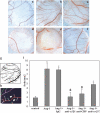
α5β1 is essential for Ang-1–mediated angiogenesis in CAM assay. Filter disks saturated with saline (a), Ang-1 (b), Ang-1 treated with control immunoglobulins (c), anti-α5β1 (JBS5; d), anti-CBP (784A2A6; e), and anti-αvβ3 (LM609; f) Abs. The vessel pattern (g) and forks (h) were analyzed by the imaging software winRHIZO Pro. (i) Measurement of forks/mm2 by the imaging software winRHIZO Pro. Values shown are means ± SD of five independent experiments, each in triplicate. Statistical significance (*, P < 0.01) is shown for Ang-1 treatment compared with control and (§, P < 0.01) for Abs treatment compared with Ang-1 stimulation.

References

    1. Arai, F., A. Hirao, M. Ohmura, H. Sato, S. Matsuoka, K. Takubo, K. Ito, G.Y. Koh, and T. Suda. 2004. Tie2/angiopoietin-1 signaling regulates hematopoietic stem cell quiescence in the bone marrow niche. Cell. 118:149–161. - PubMed
    1. Armstrong, P.B., and M.T. Armstrong. 2000. Intercellular invasion and the organizational stability of tissues: a role for fibronectin. Biochim. Biophys. Acta. 1470:9–20. - PubMed
    1. Arnaout, M.A., S.L. Goodman, and J.P. Xiong. 2002. Coming to grips with integrin binding to ligands. Curr. Opin. Cell Biol. 14:641–651. - PubMed
    1. Asahara, T., D. Chen, T. Takahashi, K. Fujikawa, M. Kearney, M. Magner, G.D. Yancopoulos, and J.M. Isner. 1998. Tie2 receptor ligands, angiopoietin-1 and angiopoietin-2, modulate VEGF-induced postnatal neovascularization. Circ. Res. 83:233–240. - PubMed
    1. Audero, E., I. Cascone, F. Maniero, L. Napione, M. Arese, L. Lanfrancone, and F. Bussolino. 2004. Adaptor ShcA protein binds tyrosine kinase Tie2 receptor and regulates migration and sprouting but not survival of endothelial cells. J. Biol. Chem. 279:13224–13233. - PubMed

Publication types

MeSH terms