. 2005 Sep;187(18):6354-62.
doi: 10.1128/JB.187.18.6354-6362.2005.

Independent and interchangeable multimerization domains of the AbrB, Abh, and SpoVT global regulatory proteins

Affiliations

Independent and interchangeable multimerization domains of the AbrB, Abh, and SpoVT global regulatory proteins

Fude Yao et al. J Bacteriol. 2005 Sep.

Erratum in

  • J Bacteriol. 2005 Nov;187(21):7546

Abstract

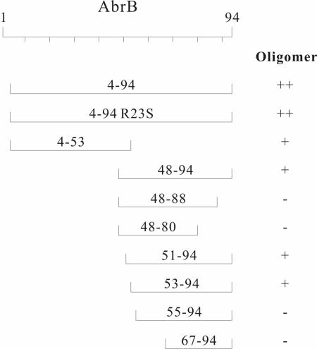
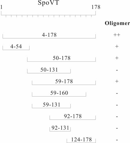
The global regulators AbrB, Abh, and SpoVT are paralogous proteins showing their most extensive sequence homologies in the DNA-binding amino-terminal regions (about 50 residues). The carboxyl-terminal portion of AbrB has been hypothesized to be a multimerization domain with little if any role in DNA-binding recognition or specificity. To investigate the multimerization potentials of the carboxyl-terminal portions of AbrB, Abh, and SpoVT we utilized an in vivo multimerization assay system based upon fusion of the domains to the DNA binding domain of the lambda cI repressor protein. The results indicate that the N and C domains of all three paralogues are independent dimerization modules and that the intact Abh and SpoVT proteins are most probably tetramers. Chimeric proteins consisting of the AbrB N-terminal DNA-binding domain fused to the C domain of either Abh or SpoVT are indistinguishable from wild-type AbrB in their ability to regulate an AbrB target promoter in vivo.


Figures

FIG. 1.
FIG. 1.
Sequence alignment of the B. subtilis AbrB, Abh, and SpoVT proteins. An asterisk indicates positions of amino acid identity in all three paralogues; a period denotes positions where two of the three proteins have identical amino acids at that position and the third protein has a related amino acid. The pound sign (#) denotes the conserved proline at position 50 in AbrB and Abh (position 51 of SpoVT).
FIG. 2.
FIG. 2.
Summary of assay results for fusion constructs present in the E. coli JH372 reporter strain. The results for each of the λcI DBD fusions we constructed and examined in the JH372 reporter strain are depicted. The extent of each segment from AbrB, Abh, and SpoVT fused to the λcI DBD is denoted numerically (e.g., 4-94 indicates fusion of amino acid residues spanning position 4 to position 94). The AbrB monomer is 94 residues in length, Abh is 92 residues, and SpoVT is 178 residues. Fused fragments containing a missense mutation are indicated by the nature of the amino acid change (one-letter code) at the residue position relative to the native, intact protein monomer (e.g., C54Y indicates a mutation changing cysteine to tyrosine at the 54th residue relative to the native monomer start point). The results of both phage immunity tests and β-galactosidase assays are given. S, sensitive to λ infection (i.e., no multimerization brought about by the fused segment); I, immune to λ infection (i.e., the fused segment can self-assemble into a multimeric state). The dimeric and tetrameric forms of the GCN4 protein fusions used as controls are described in references and . (Figures 3, 5, and 6 present cartoons summarizing different aspects of these results along with the results shown in Fig. 4.)
FIG. 3.
FIG. 3.
Summary of multimerization abilities of portions of the AbrB protein. The oligomeric natures of (λcI DBD)-(AbrB segments) fusion proteins, as determined on the basis of the results shown in Fig. 2 and 4, are indicated. +, dimer; −, monomer; ++, greater than dimers (i.e., tetramers).
FIG. 4.
FIG. 4.
Assessment of the ability of select AbrB and SpoVT domains to self-assemble into oligomer states greater than dimers. The results of β-galactosidase assays for various fusions present in the differential reporter strains XZ980 and JH607 are shown. Segments fused to the λcI DBD are denoted as given in the legend to Fig. 2. For an indicated fusion, the ratio of activity in XZ980 versus JH607 is indicated numerically. A XZ980/JH607 ratio of less than 1 is indicative of dimer formation; a ratio greater than 1 indicates formation of trimers or greater (see text for description and discussion). w.t., wild type.
FIG. 5.
FIG. 5.
Effects of single amino acid changes on the multimerization ability of the AbrB N and C domains. The oligomeric natures of (λcI DBD)-(AbrB segments containing missense mutations) fusion proteins, as determined on the basis of the results shown in Fig. 2 and 4, are indicated. +, dimer; −, monomer.
FIG. 6.
FIG. 6.
Summary of multimerization abilities of portions of the SpoVT protein. The oligomeric natures of (λcI DBD)-(SpoVT segments) fusion proteins, as determined on the basis of the results shown in Fig. 2 and 4, are indicated. +, dimer; −, monomer; ++, greater than dimers (probably tetramers).
FIG. 7.
FIG. 7.
Abh and SpoVT C domains can substitute for the AbrB C domain in vivo. Time course of β-galactosidase expression from an abrB8-lacZ reporter in strains expressing IPTG-inducible chimeric fusions of the N-terminal DNA-binding domain of AbrB to the C-terminal multimerization domains of Abh and SpoVT. Closed circles, pSpac-abrB (wild type), no IPTG; open circles, pSpac-abrB (wild type) plus 1 mM IPTG; closed squares, pSpac-abrBN-Cabh, no IPTG; open squares, pSpac-abrBN-Cabh plus 1 mM IPTG; closed triangles, pSpac-abrBN-CspoVT, no IPTG; open triangles, pSpac-abrBN-CspoVT plus 1 mM IPTG. Zero on the abscissa denotes the end of exponential growth and entry into stationary phase.

References

    1. Bagyan, I., J. Hobot, and S. Cutting. 1996. A compartmentalized regulator of developmental gene expression in Bacillus subtilis. J. Bacteriol. 178:4500-4507. - PMC - PubMed
    1. Beckett, D., D. S. Burz, G. K. Ackers, and R. T. Sauer. 1993. Isolation of lambda repressor mutants with defects in cooperative operator binding. Biochemistry 32:9073-9079. - PubMed
    1. Benson, L. M., J. L. Vaughn, M. A. Strauch, B. G. Bobay, R. Thompson, S. Naylor, and J. Cavanagh. 2002. Macromolecular assembly of the transition state regulator AbrB in its unbound and complexed states probed by microelectrospray ionization mass spectrometry. Anal. Biochem. 306:222-227. - PubMed
    1. Bobay, B. G., L. Benson, S. Naylor, B. Feeney, A. C. Clark, M. B. Goshe, M. A. Strauch, R. Thompson, and J. Cavanagh. 2004. Evaluation of the DNA binding tendencies of the transition state regulator AbrB. Biochemistry 43:16106-16118. - PubMed
    1. Branda, S. S., J. E. Gonzalez-Pastor, S. Ben Yehuda, R. Losick, and R. Kolter. 2001. Fruiting body formation by Bacillus subtilis. Proc. Natl. Acad. Sci. USA 98:11621-11626. - PMC - PubMed

Publication types

MeSH terms

LinkOut - more resources