Anoxic-epileptic seizures: observational study of epileptic seizures induced by syncopes
- PMID: 16159903
- PMCID: PMC1720208
- DOI: 10.1136/adc.2005.075408
Anoxic-epileptic seizures: observational study of epileptic seizures induced by syncopes
Abstract
Aims: To describe a large series of children with anoxic-epileptic seizures (AES)--that is, epileptic seizures induced by syncopes.
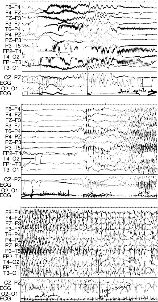
Methods: Retrospective case-note review in a tertiary paediatric neurology unit. For all 27 children seen with a definite diagnosis of AES between 1972 and 2002, a review of clinical histories, videotapes, and EEG/ECG studies was undertaken. Main outcome measures were: age of onset, frequency and type of syncopes; age of onset and frequency of AES; type and duration of induced epileptic seizures; effect of treatment of syncopal and epileptic components.
Results: Median age of onset of syncopes was 8 months (range 0.2-120), frequency 2 in total to 40/day, median total approximately 200. Syncopes were predominantly reflex asystolic (RAS), prolonged expiratory apnoea (cyanotic breath-holding spells), or of mixed or uncertain origin; there was one each of ear piercing and hair grooming vasovagal syncope and one of compulsive Valsalva. Median age of onset of AES was 17 months (range 7-120), frequency from total 1 to 3/day, median total 3. The epileptic component was almost always bilateral clonic; three had additional epilepsy, one each with complex partial seizures, myoclonic absences, and febrile seizures plus. Median duration of epileptic component was 5 minutes (range 0.5-40, mean 11). Cardiac pacing prevented RAS in two patients: most other anti-syncope therapies were ineffective. Diazepam terminated the epileptic component in 6/8. Valproate or carbamazepine abolished AES in 5/7 without influencing syncope frequency.
Conclusions: Although uncommon compared with simple syncopes, syncope triggered epileptic seizures (AES) are an important treatable basis of status epilepticus.
Figures