Review
. 2005 Aug;3(3):166-75.
doi: 10.3121/cmr.3.3.166.

Immunoregulation of dendritic cells

Affiliations
Review

Immunoregulation of dendritic cells

Mark A Wallet et al. Clin Med Res. 2005 Aug.

Abstract

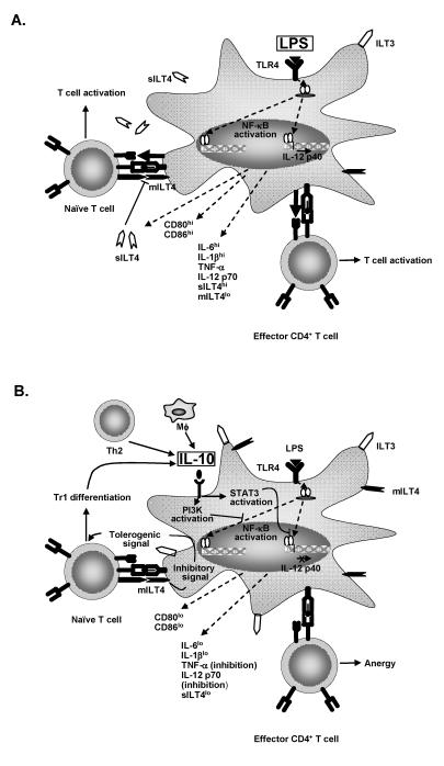
The paradigm of tolerogenic/immature versus inflammatory/mature dendritic cells has dominated the recent literature regarding the role of these antigen-presenting cells in mediating immune homeostasis or self-tolerance and response to pathogens, respectively. This issue is further complicated by the identification of distinct subtypes of dendritic cells that exhibit different antigen-presenting cell effector functions. The discovery of pathogen-associated molecular patterns and toll-like receptors provides the mechanistic basis for dendritic cell recognition of specific pathogens and induction of appropriate innate and adaptive immune responses. Only recently has insight been gained into how dendritic cells contribute to establishing and/or maintaining immunological tolerance to self. Soluble and cellular mediators have been reported to effectively regulate the function of dendritic cells by inducing several outcomes ranging from non-inflammatory dendritic cells that lack the ability to induce T lymphocyte activation to dendritic cells that actively suppress T lymphocyte responses. A thorough discussion of these stimuli and their outcomes is essential to understanding the potential for modulating dendritic cell function in the treatment of inflammatory disease conditions.


Figures

Figure 1.
Figure 1.
IL-10 induces immunoregulation of dendritic cell maturation. (A) Lipopolysaccharide promotes T cell activation. When immature dendritic cells encounter inflammatory stimuli, such as lipopolysaccharide, maturation ensues. NF-κ B is activated, translocated to the nucleus and transcription of multiple genes is induced. Costimulatory molecules CD80 and CD86, inflammatory cytokines (IL-6, IL-1β , TNF-α and IL-12p70) and soluble ILT4 (soluble LIR2) are produced which support activation of T cells. (B) IL-10 blocks the effects of lipopolysaccharide upon dendritic cells. IL-10 produced by monocytes, TH2 cells and Tr1 cells induces PI3K and signal transducer and activator of transcription (STAT)-3 activation by dendritic cells. PI3K activation results in blockade of NF-κ B activation and STAT-3 blocks binding of NF-κ B to the IL-12p40 promoter. Consequently, differentiation of Th1 T lymphocytes is inhibited. Additionally, IL-10 inhibits production of soluble ILT4 favoring expression of membrane bound ILT4. Ligation of dendritic cell membrane bound ILT4 by T lymphocytes induces a regulatory dendritic cell phenotype that induces Tr1 differentiation.
Figure 2.
Figure 2.
TGF-β 1 modulated dendritic cell phenotype and T lymphocyte activation. TGF-β 1 is produced by monocytes or macrophages, T lymphocytes or granulocytes, or is provided exogenously as a means of immunotherapy. Dendritic cells that encounter TGF-β 1 down-regulate expression of CD80, CD86, TLR4 and, importantly, CCR7. Low expression of CCR7 inhibits MIP-3β-induced migration to lymph nodes. Therefore, T lymphocyte stimulation is inhibited.
Figure 3.
Figure 3.
Apoptotic cells induce immunoregulation and tolerance induction through dendritic cells. Dendritic cells scavenge tissues for microbes and dying tissue. Apoptotic cells induce PI3K dependent signaling resulting in inhibition of NF-κ B translocation and costimulatory molecule (CD80/86) expression. Production of IL-6, TGF-β and IL-12p40 ensue. IL-12p40 homodimers inhibit the effects of IL-12p70 upon T lymphocytes, effectively inhibiting Th1 induction. Apoptotic cells induce a lasting inhibitory effect which renders dendritic cells refractory to lipopolysaccharide-induced stimulation of Th1 lymphocytes.
Figure 4.
Figure 4.
CTLA-4 and CD28 induce divergent outcomes in dendritic cell maturation. Regulatory T lymphocyte cells expressing CTLA-4 ligate CD80 on dendritic cells inducing STAT-1 activation, IFN-γ and IDO production. IDO catalyzes the breakdown of tryptophan resulting in T lymphocyte activation followed by rapid induction of apoptosis. In contrast, activated T lymphocytes expressing CD28 induce activational signals through CD80/CD86. Receptor ligation induces p38 MAPK and NF-κ B activation. IL-6 and IFN-γ are then produced. IL-6 induces a dominant antagonistic effect upon IDO production. Dendritic cells are now capable of inducing T lymphocyte activation and proliferation.

References

    1. Alaniz RC, Sandall S, Thomas EK, Wilson CB. Increased dendritic cell numbers impair protective immunity to intracellular bacteria despite augmenting antigen-specific CD8+ T lymphocyte responses. J Immunol 2004;172:3725–3735. - PubMed
    1. Maldonado-Lopez R, Moser M. Dendritic cell subsets and the regulation of Th1/Th2 responses. Semin Immunol 2001;13:275–282. - PubMed
    1. Eisenbarth SC, Piggott DA, Bottomly K. The master regulators of allergic inflammation: dendritic cells in Th2 sensitization. Curr Opin Immunol 2003;15:620–626. - PubMed
    1. Smith CM, Wilson NS, Waithman J, Villadangos JA, Carbone FR, Heath WR, Belz GT. Cognate CD4(+) T cell licensing of dendritic cells in CD8(+) T cell immunity. Nat Immunol 2004;5:1143–1148. - PubMed
    1. Gerloni M, Lo D, Zanetti M. DNA immunization in relB-deficient mice discloses a role for dendritic cells in IgM—>IgG1 switch in vivo. Eur J Immunol 1998;28:516–524. - PubMed

Substances

LinkOut - more resources