. 2005 Oct;79(19):12205-17.
doi: 10.1128/JVI.79.19.12205-12217.2005.

Mitochondrial cell death suppressors carried by human and murine cytomegalovirus confer resistance to proteasome inhibitor-induced apoptosis

Affiliations

Mitochondrial cell death suppressors carried by human and murine cytomegalovirus confer resistance to proteasome inhibitor-induced apoptosis

A Louise McCormick et al. J Virol. 2005 Oct.

Abstract

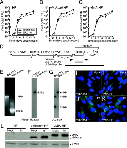
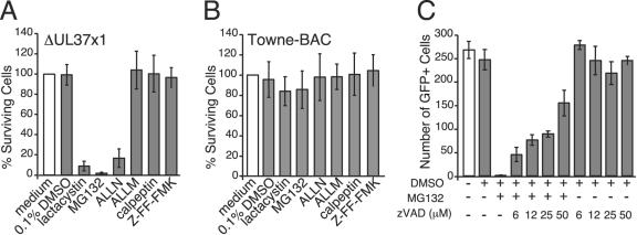
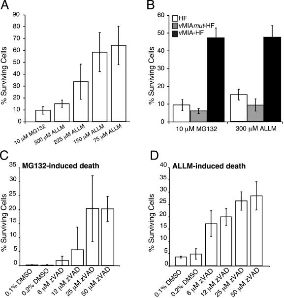
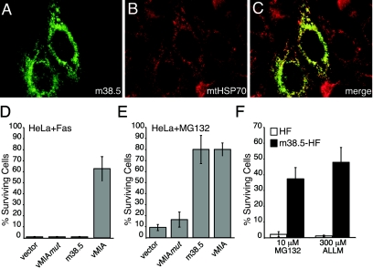
Human cytomegalovirus carries a mitochondria-localized inhibitor of apoptosis (vMIA) that is conserved in primate cytomegaloviruses. We find that inactivating mutations within UL37x1, which encodes vMIA, do not substantially affect replication in TownevarATCC (Towne-BAC), a virus that carries a functional copy of the betaherpesvirus-conserved viral inhibitor of caspase 8 activation, the UL36 gene product. In Towne-BAC infection, vMIA reduces susceptibility of infected cells to intrinsic death induced by proteasome inhibition. vMIA is sufficient to confer resistance to proteasome inhibition when expressed independent of viral infection. Murine cytomegalovirus m38.5, whose position in the viral genome is analogous to UL37x1, exhibits mitochondrial association and functions in much the same manner as vMIA in inhibiting intrinsic cell death. This work suggests a common role for vMIA in rodent and primate cytomegaloviruses, modulating the threshold of virus-infected cells to intrinsic cell death.


Figures

FIG. 1.
FIG. 1.
ΔUL37x1 virus yield and complementation. (A to C) Mean viral yields from triplicate cultures of HFs (A), vMIAmut-HFs (B), or vMIA-HFs (C) infected at an MOI of 0.002 by ΔUL37x1 or Towne-BAC, graphed with standard deviations (hidden behind the symbols in most cases). (D) Map of the UL36-39 region showing positions of oligonucleotide primers (arrows) and probes (thick lines) used in characterizing the ΔUL37x1 virus genome structure. The ORF map shows splicing patterns, transcript orientation (arrowheads), and the position of the KanMX4 cassette replacement of UL37x1 (dashed lines). (E) UL37x1 region PCR products from ΔUL37x1 or Towne-BAC viral DNA, as indicated, resolved by electrophoresis in agarose gels and visualized by ethidium bromide fluorescence. (F and G) Autoradiographs of EcoRI-digested ΔUL37x1 or Towne-BAC viral DNAs hybridized with UL37x1 probe (F) or UL38-39 probe, consistent with the expected additional EcoRI site due to the KanMX4 cassette in the mutant (G). (H to K) IFA of gene expression in vMIAmut-HF (H and J) or vMIA-HF (I and K) without infection (mock; H to I) or following Towne-BAC infection at an MOI of 3 for 24 h (Inf; J to K). Shown is a composite of vMIAmut and vMIA proteins (green) and nuclei (blue). (L) Immunoblot analysis of vMIAmut and vMIA in lysates of HFs, vMIAmut-HFs, and vMIA-HFs following infection (Inf) by Towne-BAC at an MOI of 3 for 24 h, as indicated. Shown are vMIAmut and vMIA proteins detected with anti-Myc antibody (upper panel) along with endogenous c-Myc protein (lower panel) as a loading control.
FIG. 2.
FIG. 2.
Growth and genomic structure properties of ΔUL37x1 viruses derived on complementing vMIA-HF and noncomplementing HF cells. (A) Mean viral yields with standard deviations from HF cultures infected for 14 days with three transfection-independent viruses derived on HF or vMIA-HF at an MOI of 0.001. (B) Restriction fragment length polymorphism analysis of EcoRV-digested ΔUL37x1 or Towne-BAC virion DNA recovered from independent transfections of HFs (isolates a, b, and c) and vMIA-HFs (isolates d, e, and f), with two dashes indicating the expected polymorphism due to insertion of the KanMX4 cassette. (C and D) Infected cell and focus formation by ΔUL37x1 and Towne-BAC following transfection of equivalent amounts of BAC DNA into noncomplementing HFs. The mean numbers of single GFP-positive (GFP+) cells (C) and GFP-positive foci (D), monitored by live cell microscopy, are graphed with standard deviations from four independent transfections of ΔUL37x1 or Towne-BAC.
FIG. 3.
FIG. 3.
Cell death and vMIA suppression in ΔUL37x1 cultures. (A and B) Fluorescence images of GFP expression on day 10 pi of a ΔUL37x1 (A) or Towne-BAC (B) plaque. Arrowheads indicate fragmented cells. (C and D) ΔUL37x1 (GFP+) infected cell death frequency (fragmented cells) at 72 hpi on HF, vMIAmut-HF, or vMIA-HF (C) and on HFs exposed to zVAD (D), as indicated. Fragmented cells from four independent cultures were counted and are graphed as the mean number of fragmented cells relative to total GFP+ cells (percent fragmented GFP+ cells), with standard deviations indicated by error bars. Percent fragmentation was determined from >1,000 infected cells in total. (E) Impact of zVAD as indicated on day 7 viral yields of ΔUL37x1 or Towne-BAC.
FIG. 4.
FIG. 4.
Protease inhibitor-induced cell death of ΔUL37x1-infected HFs. HFs were infected with ΔUL37x1 (A, C, and E to J) or Towne-BAC (B, D, and K to P) for 48 h, before replacement of medium with normal medium (A, B, E, and K), medium plus 0.1% DMSO (F and L), or medium including 0.1% DMSO and 100 μM ALLM (C, D, I, and O), 100 μM Z-FF-FMK (G and M), 100 μM calpeptin (H and N), or 10 μM MG132 (J and P). Images are from GFP fluorescence 20 h post-media replacement. Arrowheads are placed at examples of fragmented cells in ΔUL37x1-infected cultures (A and C).
FIG. 5.
FIG. 5.
Caspase-dependent death induced in ΔUL37x1-infected cells by 10 μM proteasome inhibitors. (A and B) HFs infected with ΔUL37x1 (A) or Towne-BAC (B) for 48 h were exposed to 10 μM concentrations of lactacystin, MG132, ALLN, ALLM, calpeptin, or Z-FF-FMK for 20 h in medium containing 0.1% DMSO, control medium with 0.1% DMSO), or normal medium without DMSO (open bar). Surviving cells from three independent experiments were counted and are graphed as the mean percent survival relative to the mean number of viable cells in the medium-only cultures (percent surviving cells), with standard deviations indicated by error bars. Percent survival was determined from >1,000 infected cells per inhibitor. (C) HFs infected with ΔUL37x1 virus were exposed for 20 h to 10 μM MG132, zVAD at the concentrations indicated, control medium (open bar), or medium plus 0.1% DMSO. Graphed are the mean numbers of viable GFP-positive cells determined in three separate cultures, with error bars indicating the standard deviations between cultures.
FIG. 6.
FIG. 6.
Kinetics, dose dependence, and protection by vMIA of death induced by proteasome inhibition. (A and B) HFs, vMIAmut-HFs, and vMIA-HF were exposed to 10 μM MG132 or ALLM at 300 μM, 225 μM, 150 μM, or 75 μM as indicated. At 44 h postaddition, surviving cells from five microscopic fields were counted and are graphed as the mean percent survival relative to controls, with standard deviations indicated by error bars. Each field of untreated control cells included approximately 1,000 cells. (C and D) HFs were exposed to 10 μM MG132 (C) or 300 μM ALLM (D) in the presence or absence of zVAD at the concentrations indicated. A 0.1% DMSO concentration was equivalent to the solvent from proteasome inhibitors, and 0.2% DMSO was equivalent to the final concentration in all cultures that received proteasome inhibitor plus zVAD at the concentrations indicated. Analysis followed as for panels A and B.
FIG. 7.
FIG. 7.
Subcellular localization of m38.5 and function in cell death assays. (A to C) m38.5 (green) (A), detected with antibodies to the myc epitope, and mitochondria (red) (B), detected with antibodies to mtHSP70, 24 h posttransfection of HeLa cells with LNCX-m38.5. Individual images were merged for panel C. (D and E) Survival of HeLa cells transfected with LNCX-GFP and LNCX-3myc (vector), LNCX-vMIAmut, LNCX-m38.5, or LNCX-vMIA exposed to anti-Fas antibody plus CH or MG132 added to culture wells for 20 h (Fas) or 44 h (MG132). Surviving GFP-positive cells from three independent transfections (nine fields) were counted and graphed as described for Fig. 6. Each field of untreated cells included an average of 90 GFP-positive cells. (F) m38.5-HFs or control HFs were exposed to 10 μM MG132 or 300 μM ALLM for 44 h. Surviving cells from five fields were counted and graphed as described for Fig. 6. Each field of untreated cells included an average of 1,000 cells.

Similar articles

Cited by

References

    1. Alcami, A., and U. H. Koszinowski. 2000. Viral mechanisms of immune evasion. Trends Microbiol. 8:410-418. - PMC - PubMed
    1. Andoniou, C., D. Andrews, M. Manzur, P. Ricciardi-Castagnoli, and M. Degli-Esposti. 2004. A novel checkpoint in the Bcl-2-regulated apoptotic pathway revealed by murine cytomegalovirus infection of dendritic cells. J. Cell Biol. 166:827-837. - PMC - PubMed
    1. Arnoult, D., L. Bartle, A. Skaletskaya, D. Poncet, N. Zamzami, P. Park, J. Sharpe, R. Youle, and V. S. Goldmacher. 2004. Cytomegalovirus cell death suppressor vMIA blocks Bax- but not Bak-mediated apoptosis by binding and sequestering Bax at mitochondria. Proc. Natl. Acad. Sci. USA 101:7988-7993. - PMC - PubMed
    1. Baldick, C. J., Jr., A. Marchini, C. E. Patterson, and T. Shenk. 1997. Human cytomegalovirus tegument protein pp71 (ppUL82) enhances the infectivity of viral DNA and accelerates the infectious cycle. J. Virol. 71:4400-4408. - PMC - PubMed
    1. Bartz, S., and M. Vodicka. 1997. Production of high-titer human immunodeficiency virus type 1 pseudotyped with vesicular stomatitis virus glycoprotein. Methods 12:337-342. - PubMed

Publication types

MeSH terms