. 2005 Oct;79(19):12218-30.
doi: 10.1128/JVI.79.19.12218-12230.2005.

Four-dimensional visualization of the simultaneous activity of alternative adeno-associated virus replication origins

Affiliations

Four-dimensional visualization of the simultaneous activity of alternative adeno-associated virus replication origins

Daniel L Glauser et al. J Virol. 2005 Oct.

Abstract

The adeno-associated virus (AAV) inverted terminal repeats (ITRs) contain the AAV Rep protein-binding site (RBS) and the terminal resolution site (TRS), which together act as a minimal origin of DNA replication. The AAV p5 promoter also contains an RBS, which is involved in Rep-mediated regulation of promoter activity, as well as a functional TRS, and origin activity of these signals has in fact been demonstrated previously in the presence of adenovirus helper functions. Here, we show that in the presence of herpes simplex virus type 1 (HSV-1) and AAV Rep protein, p5 promoter-bearing plasmids are efficiently amplified to form large head-to-tail concatemers, which are readily packaged in HSV-1 virions if an HSV-1 DNA-packaging/cleavage signal is provided in cis. We also demonstrate simultaneous and independent replication from the two alternative AAV replication origins, p5 and ITR, on the single-cell level using multicolor-fluorescence live imaging, a finding which raises the possibility that both origins may contribute to the AAV life cycle. Furthermore, we assess the differential affinities of Rep for the two different replication origins, p5 and ITR, both in vitro and in live cells and identify this as a potential mechanism to control the replicative and promoter activities of p5.


Figures
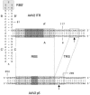
FIG. 1.
FIG. 1.
Sequence alignment of the RBS and TRS of the AAV2 ITR and the AAV2 p5 promoter. The 25-bp RBS of the A-stem (28) and its homologue in the p5 promoter (28) are indicated by boxes in light gray. The A-stem RBS contains three perfect GAGC tetranucleotide repeats (dark-gray boxes) flanked by two imperfect repeats, while the p5 RBS contains only one perfect GAGC repeat and four imperfect repeats. The CTTTG element within the secondary-structure element of the ITR, which was previously described as enhancing Rep binding affinity (41), is indicated by a light-gray box and is termed RBS′. The TRS of the A-stem (2, 17) and its homologue in the p5 promoter (54) are indicated by open boxes. Nicking at the A-stem TRS occurs between the two thymidine bases (top arrow), while at the p5 promoter TRS it occurs between a thymidine and an adenosine base (bottom arrow). Note that the p5 TRS is spaced 10 bp closer to the RBS than the A-stem TRS. The borders of the DNA substrates used in the EMS assays (Fig. 8 and 9) are indicated by the numbered AAV2 nucleotides: ITR substrate, nt 9 to 117 (single stranded and self-annealed); A-stem substrate, nt 81 to 117 (double stranded); p5 substrate, nt 254 to 290 (double stranded).
FIG. 2.
FIG. 2.
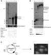
Replication of AAV p5-containing plasmids. (A) Replication of plasmid pRep-red. VERO 2-2 cells were cotransfected with pRep (lanes 1 to 6) and either pRep-red (lanes 1 to 4) or pCMVrep-red (lanes 5 and 6). Subsequently, the cells were superinfected with HSV-1 (lanes 1, 2, 5, and 6). Hirt DNA prepared 48 h later was digested with DpnI and/or BglII and analyzed by Southern blotting with a DIG-labeled DsRed probe. The sizes of the linearized plasmids pRep-red (5.9 kb) and pCMVrep-red (6.2 kb), as well as a prominent fragment of the DpnI digest, are indicated (arrows). (B) Conformation of p5 replication products. VERO 2-2 cells were transfected with plasmid pRep and superinfected with HSV-1. Hirt DNA was prepared as in panel A, digested with DpnI and decreasing amounts of BglII, and analyzed by Southern blotting with a DIG-labeled rep probe. The bands corresponding to the linear monomer and multimers of pRep and a prominent fragment of the DpnI digest are indicated (arrows). The fragment sizes of the DIG-labeled molecular weight standard (M) are indicated on the left. +, present; −, absent. (C) Packaging of p5 replication products into HSV-1 particles. Schematic representation of pP5cagGFP (left) and representative fluorescence micrograph of transduced cells (right). The AAV p5 promoter (AAV p5), the HSV-1 DNA-packaging/cleavage signal (HSV-1 pac), the rep-ecfp fusion gene (rep-ecfp), and the EGFP expression cassette (cag-egfp) are shown.
FIG. 3.
FIG. 3.
Live visualization of AAV DNA replication from alternative replication origins. (A) Schematic representation of live-visualization assays. Replication from the AAV p5 ori is visualized with plasmid pBs-p5-tetO, which contains the AAV p5 promoter (p5) and five reiterations of the seven-copy tetO sequence comprising a total of 35 TetR binding sites. HSV-1 and Rep-dependent accumulation of concatameric replication products (HMW) is visualized by binding of an ECFP-TetR fusion protein. Visualization of replication from the ITR origins (pAAVlacO), which employs lacO interactions with an EYFP-LacI fusion, has been described previously (9). Double-stranded monomeric (ITRm) and dimeric (ITRd) replication intermediates are indicated. (B) Replication assay with plasmid pBs-p5-tetO. VERO 2-2 cells were cotransfected with pBs-p5-tetO and pCMVrep68/78-kan (lanes 1 and 4) or pCMVrep-HcRed (lane 2); pBs-p5-tetO (lane 3); or pBs-tetO and pCMVrep68/78-kan (lane 5) and superinfected with HSV-1 (lanes 1, 2, 3, and 5). Hirt DNA was prepared at 48 h p.i., digested with DpnI, and analyzed by Southern blotting with a probe for the ampicillin resistance (Ampr) gene present on pBs-p5-tetO. Concatameric, high-molecular-weight replication products are indicated (HMW). +, present; −, absent. (C) Formation of nuclear AAV p5 replication compartments in the presence of the p5 ori, AAV rep, and HSV-1. Cells were cotransfected with pBs-p5-tetO, pEYFPTetR, and pCMVrep68/78-kan (a and c), pBs-p5-tetO and pEYFPTetR (b), or pBs-tetO, pEYFPTetR, and pCMVrep68/78-kan (d) and superinfected with HSV-1 (a, b, and d) or mock infected (c). The micrographs were taken 36 h p.i. under a standard fluorescence microscope with a filter specific for EYFP.
FIG. 4.
FIG. 4.
Formation of AAV p5 replication compartments in live HeLa cells. Cells were cotransfected with pBs-p5-tetO, pEYFPTetR, and pCMVrep-HcRed and superinfected with HSV-1. Time lapse images starting at 27.5 h p.i. were taken with a confocal microscope with settings specific for EYFP (p5 replication compartments) and HcRed (AAV Rep; inset).
FIG. 5.
FIG. 5.
Visualization of AAV p5 and ITR replication compartments (green) and AAV Rep (red) in live HeLa cells. Cells were cotransfected with pBs-p5-tetO, pEYFPTetR, and pCMVrep-HcRed (a to f) or pAAVlacO, pSV2-EYFP/lacI, and pCMVrep-HcRed (g and h). After transfection, the cells were superinfected with HSV-1. The images were taken between 24 and 36 h p.i. with a standard fluorescence microscope and filters specific for EYFP (AAV replication compartments) or Texas Red (AAV Rep). Three different nuclei containing p5 replication compartments and one nucleus containing ITR replication compartments are shown. Repl. Comp., replication compartments.
FIG.6.
FIG.6.
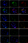
Covisualization of AAV DNA replication from alternative replication origins and AAV Rep protein. Three individual nuclei are shown. HeLa cells were cotransfected with pBs-p5-tetO, pAAVlacO, pECFPTetR, pSV2-EYFP/lacI, and pCMVrep-HcRed and superinfected with HSV-1. Images of live (a to f) or fixed (g to s) cells were taken between 36 and 48 h p.i. with a confocal microscope with settings specific for ECFP (p5 replication compartments), EYFP (ITR replication compartments), and HcRed (Rep protein). The images represent projections through three-dimensional reconstructions of nuclei.
FIG. 7.
FIG. 7.
Three-dimensional view of the nucleus presented in Fig. 6 (a to f). p5 replication compartments are stained blue, ITR replication compartments green, and Rep protein red. Deconvolved three-dimensional reconstructions of the nucleus were processed in Imaris software using the surpass view mode.
FIG. 8.
FIG. 8.
Rep binding to AAV replication origins: titration of Rep protein. (A) Twofold dilutions of Rep protein were bound to 0.04 pmol of radiolabeled DNA substrates consisting of the hairpinned ITR (ITR), the linear A-stem (A-stem), or the linear p5 substrate (p5). (B) Quantification of the fraction of bound DNA substrate. The PhosphorImager data were analyzed by ImageQuant software, and the bound fraction (y axis) was plotted against the amount of Rep protein (x axis).
FIG. 9.
FIG. 9.
Rep binding to AAV replication origins: reciprocal competition analysis. Fifteen nanograms (A and B) or 60 ng (C) of Rep protein was bound to 0.1 pmol of radiolabeled ITR (A), A-stem (B), or p5 DNA (C) substrate in the presence of a 0.1-, 1-, 10-, or 100-fold amount of unlabeled ITR, A-stem, or p5 competitor DNA. The diagrams at the bottom represent the relative binding (y axis) as a function of the amount (n-fold) of competitor DNA (fold competitor; x axis). The amount of bound probe was quantified by ImageQuant analysis of the PhosphoImager data, and the value in the absence of competitor DNA was set as 100%. Comp., Competitor; FP, free probe; NC, no competitor.

References

    1. Balague, C., M. Kalla, and W. W. Zhang. 1997. Adeno-associated virus Rep78 protein and terminal repeats enhance integration of DNA sequences into the cellular genome. J. Virol. 71:3299-3306. - PMC - PubMed
    1. Brister, J. R., and N. Muzyczka. 1999. Rep-mediated nicking of the adeno-associated virus origin requires two biochemical activities, DNA helicase activity and transesterification. J. Virol. 73:9325-9336. - PMC - PubMed
    1. Caleo, M., M. C. Cenni, M. Costa, E. Menna, L. Zentilin, S. Giadrossi, M. Giacca, and L. Maffei. 2002. Expression of BCL-2 via adeno-associated virus vectors rescues thalamic neurons after visual cortex lesion in the adult rat. Eur. J. Neurosci. 15:1271-1277. - PubMed
    1. Chadeuf, G., D. Favre, J. Tessier, N. Provost, P. Nony, J. Kleinschmidt, P. Moullier, and A. Salvetti. 2000. Efficient recombinant adeno-associated virus production by a stable rep-cap HeLa cell line correlates with adenovirus-induced amplification of the integrated rep-cap genome. J. Gene Med. 2:260-268. - PubMed
    1. Chang, L. S., Y. Shi, and T. Shenk. 1989. Adeno-associated virus P5 promoter contains an adenovirus E1A-inducible element and a binding site for the major late transcription factor. J. Virol. 63:3479-3488. - PMC - PubMed

Publication types

LinkOut - more resources