. 2005 Oct;20(10):1858-66.
doi: 10.1359/JBMR.050611. Epub 2005 Jun 27.

RhoA and cytoskeletal disruption mediate reduced osteoblastogenesis and enhanced adipogenesis of human mesenchymal stem cells in modeled microgravity

Affiliations

RhoA and cytoskeletal disruption mediate reduced osteoblastogenesis and enhanced adipogenesis of human mesenchymal stem cells in modeled microgravity

Valerie E Meyers et al. J Bone Miner Res. 2005 Oct.

Abstract

Spaceflight, aging, and disuse lead to reduced BMD. This study shows that overexpression of constitutively active RhoA restores actin cytoskeletal arrangement, enhances the osteoblastic phenotype, and suppresses the adipocytic phenotype of human mesenchymal stem cells cultured in modeled microgravity.

Introduction: Reduced BMD during spaceflight is partly caused by reduced bone formation. However, mechanisms responsible for this bone loss remain unclear. We have previously shown reduced osteoblastogenesis and enhanced adipogenesis of human mesenchymal stem cells (hMSCs) cultured in modeled microgravity (MMG). The small GTPase, RhoA, regulates actin stress fiber formation and has been implicated in the lineage commitment of hMSCs. We examined the effects of MMG on actin cytoskeletal organization and RhoA activity and the ability of constitutively active RhoA to reverse these effects.

Materials and methods: hMSCs were seeded onto plastic microcarrier beads at a density of 10(6) and allowed to form aggregates in DMEM containing 10% FBS for 7 days. Aggregates were incubated in DMEM containing 2% FBS for 6 h with or without an adenoviral vector containing constitutively active RhoA at a multiplicity of infection (moi) of 500 and allowed to recover in 10% FBS for 24 h. Cells were transferred to the rotary cell culture system to model microgravity or to be maintained at normal gravity for 7 days in DMEM, 10% FBS, 10 nM dexamethasone, 10 mM beta-glycerol phosphate, and 50 muM ascorbic acid 2-phosphate.

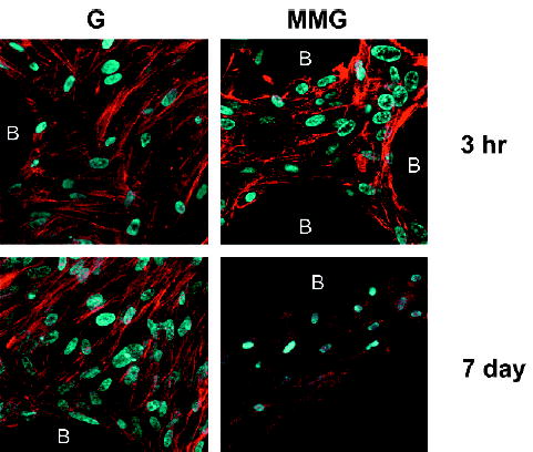
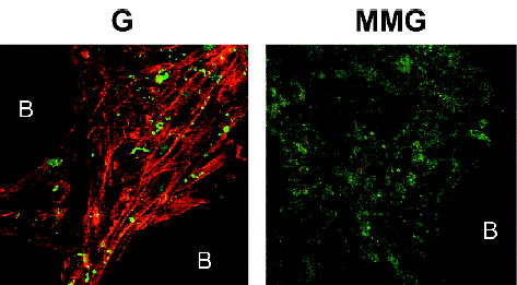
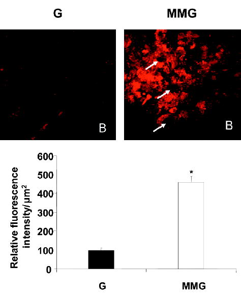
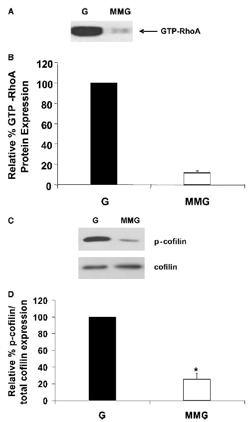
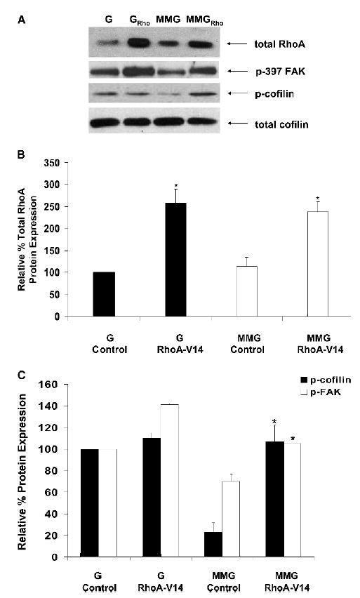
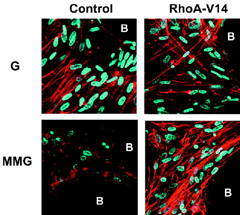
Results: F-actin stress fibers are disrupted in hMSCs within 3 h of initiation of MMG and are completely absent by 7 days, whereas monomeric G-actin is increased. Because of the association of G-actin with lipid droplets in fat cells, the observed 310% increase in intracellular lipid accumulation in hMSCs cultured in MMG was not unexpected. Consistent with these changes in cellular morphology, 7 days of MMG significantly reduces RhoA activity and subsequent phosphorylation of cofilin by 88+/-2% and 77+/-9%, respectively. Importantly, introduction of an adenoviral construct expressing constitutively active RhoA reverses the elimination of stress fibers, significantly increases osteoblastic gene expression of type I collagen, alkaline phosphatase, and runt-related transcription factor 2, and suppresses adipocytic gene expression of leptin and glucose transporter 4 in hMSCs cultured in MMG.

Conclusion: Suppression of RhoA activity during MMG represents a novel mechanism for reduced osteoblastogenesis and enhanced adipogenesis of hMSCs.


Conflict of interest statement

The authors have no conflict of interest.

Figures

FIG. 1
FIG. 1
Actin stress fiber formation is disrupted in MMG. hMSCs aggregated on plastic microcarrier beads (B) were maintained in osteogenic medium for the indicated times in normal gravity (G) or MMG. Cells were labeled with Alexa Fluor 594-conjugated phalloidin to visualize filamentous actin (red) and Hoechst stain to visualize nuclei (blue) at the end of the study. Images were acquired using laser scanning confocal microscopy under a ×63 objective. Images are representative of six separate experiments.
FIG. 2
FIG. 2
hMSCs cultured in MMG predominantly express the globular form of actin. hMSCs aggregated on plastic microcarrier beads (B) were maintained in osteogenic medium for 7 days in normal gravity (G) or MMG. Cells were double labeled with Alexa Fluor 488–conjugated deoxyribonuclease I (green) to visualize globular actin and Alexa Fluor 594–conjugated phalloidin (red) to visualize filamentous actin. Images were acquired using laser scanning confocal microscopy under a ×63 objective. Images are representative of three separate experiments.
FIG. 3
FIG. 3
MMG enhances intracellular lipid accumulation. hMSCs aggregated on plastic microcarrier beads (B) were maintained in osteogenic medium for 7 days in normal gravity (G) or MMG. Cells were stained with Nile Red to visualize cellular lipid content (red). (A) Images from three separate experiments were acquired using laser scanning confocal microscopy under a ×63 objective, and representative images are shown. White arrows indicate cell nuclei. (B) Image pixel intensity was quantified for three separate experiments using BioQuant image analysis software and are graphed as the mean ± SE; *p ≤ 0.01 compared with G.
FIG. 4
FIG. 4
RhoA activity and cofilin phosphorylation are reduced in MMG. hMSCs aggregated on plastic microcarrier beads were maintained in osteogenic medium for 7 days in normal gravity (G) or MMG. Whole cell protein was extracted at the end of the study. (A) Activated (GTP-bound) RhoA was pulled down from total protein (300 μg) using Rhotekin-conjugated agarose beads and separated by 12.5% SDS-PAGE. Immunoblots were probed using antibodies directed against RhoA. The image is representative of two independent experiments each performed in duplicate. (B) The band intensities of GTP-RhoA in MMG are graphed as a percentage relative to gravity controls (G). Values were obtained from twoseparate experiments performed in duplicate and represent the mean ± SE. (C) Total protein was separated by 12.5% SDS-PAGE. Immunoblots were probed using antibodies directed against cofilin or phosphorylated cofilin. The image is representative of five separate experiments. (D) The band intensities of phosphorylated cofilin relative to total cofilin in MMG are graphed as a percentage relative to gravity controls (G). Values were obtained from five separate experiments and represent the mean ± SE. *p ≤ 0.05 compared with G.
FIG. 5
FIG. 5
Overexpression of constitutively active RhoA induces cofilin and FAK phosphorylation in MMG. hMSCs aggregated on plastic microcarrier beads were mock-infected (Control) or infected with an adenoviral vector containing constitutively active RhoA (RhoA-V14) and were maintained in osteogenic medium for 7 days in normal gravity (G) or MMG. (A) Total protein was extracted at the end of the study and separated by SDS-PAGE. Immunoblots were probed using antibodies directed against RhoA, phosphorylated cofilin, FAK, and total cofilin. Images are representative of three separate experiments. (B) The band intensities of RhoA are graphed relative to uninfected gravity controls. Values were obtained from three separate experiments. *p ≤ 0.05 compared with controls. (C) The band intensities of phosphorylated protein relative to total protein are graphed as a percentage relative to uninfected gravity controls. Values were obtained from three separate experiments and represent the mean ± SE. *p ≤ 0.05 compared with MMG control.
FIG. 6
FIG. 6
Overexpression of constitutively active RhoA prevents loss of stress fibers in MMG. hMSCs aggregated on plastic microcarrier beads were mock-infected (Control) or infected with an adenoviral vector containing constitutively active RhoA (RhoA-V14) and were maintained in osteogenic medium for 7 days in normal gravity (G) or MMG. At the end of the study, cells were labeled with Alexa Fluor 594-conjugated phalloidin to visualize filamentous actin (red) and Hoechst stain to visualize nuclei (blue). Images were acquired by laser scanning confocal microscopy under a ×63 objective. Images are representative of three separate experiments.
FIG. 7
FIG. 7
Constitutively active RhoA induces gene expression of markers of osteoblastic differentiation. hMSCs aggregated on plastic microcarrier beads were mock infected (Control) or infected with an adenoviral vector containing constitutively active RhoA (RhoA-V14) and were maintained in osteogenic medium for 7 days in normal gravity (G) or MMG. Total RNA was extracted at the end of the study. (A) Semiquantitative RT-PCR reactions were performed using primers for osteoblastic genes, including alkaline phosphatase (ALP), preprocollagen type I (Col I), and runt-related transcription factor 2 (Runx2). Relative gene expression from three experiments was normalized to 18S expression. Values represent the mean ± SE. *p ≤ 0.05 compared with MMG control. (B) Semiquantitative RT-PCR reactions were performed using primers for the adipocytic gene, leptin (Lep). Relative gene expression from three experiments was normalized to 18S expression. Values represent the mean ± SE. *p ≤ 0.05 compared with MMG control. (C) Conventional RT-PCR reactions were performed using primers for the adipocytic gene, glucose transporter 4 (GLUT4). Images are representative of three separate experiments.

References

    1. Lang T, LeBlanc A, Evans H, Lu Y, Genant H, Yu A. Cortical and trabecular bone mineral loss from the spine and hip in long-duration spaceflight. J Bone Miner Res. 2004;19:1006–1012. - PubMed
    1. Ding M, Odgaard A, Linde F, Hvid I. Age-related variations in the microstructure of human tibial cancellous bone. J Orthop Res. 2002;20:615–621. - PubMed
    1. Giangregorio L, Blimkie CJ. Skeletal adaptations to alterations in weight-bearing activity: A comparison of models of disuse osteoporosis. Sports Med. 2002;32:459–476. - PubMed
    1. Bikle DD, Halloran BP. The response of bone to unloading. J Bone Miner Metab. 1999;17:233–244. - PubMed
    1. Vico L, Collet P, Guignandon A, Lafage-Proust MH, Thomas T, Rehaillia M, Alexandre C. Effects of long-term microgravity exposure on cancellous and cortical weight-bearing bones of cosmonauts. Lancet. 2000;355:1607–1611. - PubMed

Publication types

Substances