Clinical Trial
. 2005 Oct;2(10):e265.
doi: 10.1371/journal.pmed.0020265. Epub 2005 Sep 20.

Marked differences in human melanoma antigen-specific T cell responsiveness after vaccination using a functional microarray

Affiliations
Clinical Trial

Marked differences in human melanoma antigen-specific T cell responsiveness after vaccination using a functional microarray

Daniel S Chen et al. PLoS Med. 2005 Oct.

Abstract

Background: In contrast to many animal model studies, immunotherapeutic trials in humans suffering from cancer invariably result in a broad range of outcomes, from long-lasting remissions to no discernable effect.

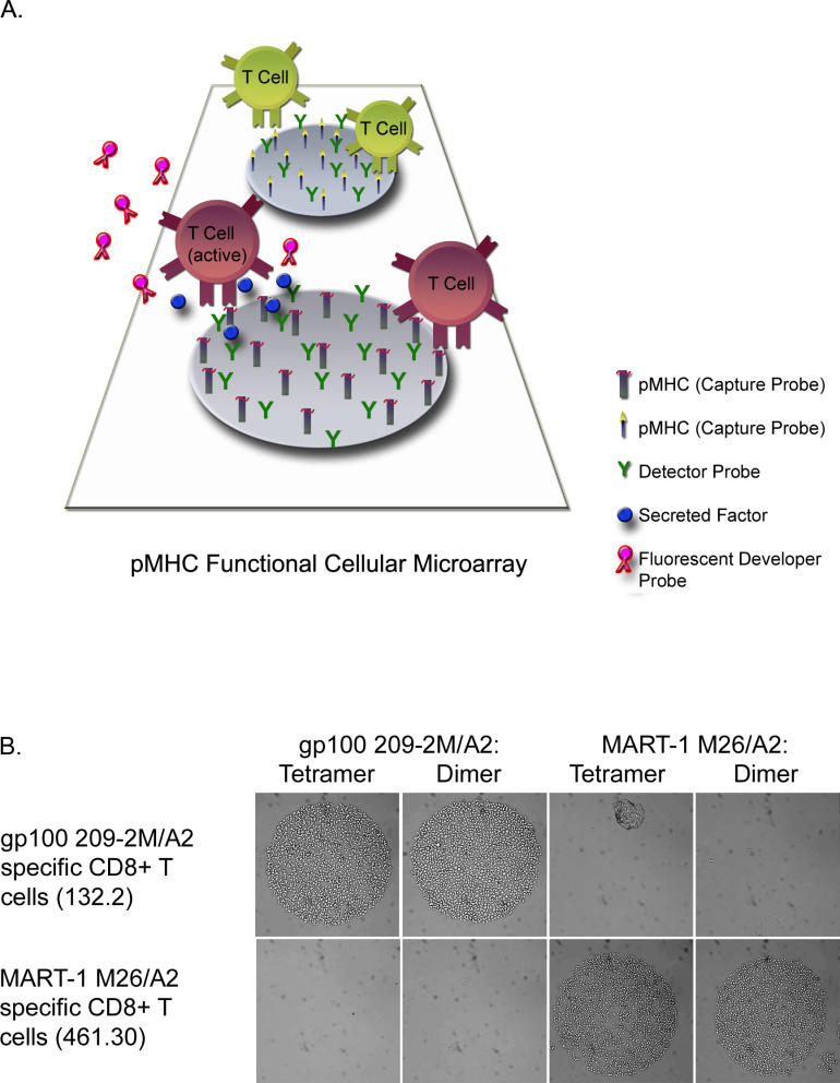
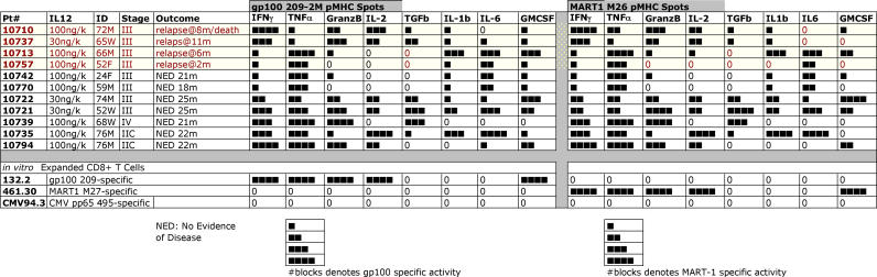
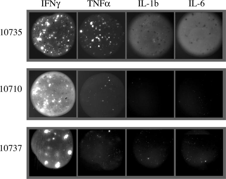
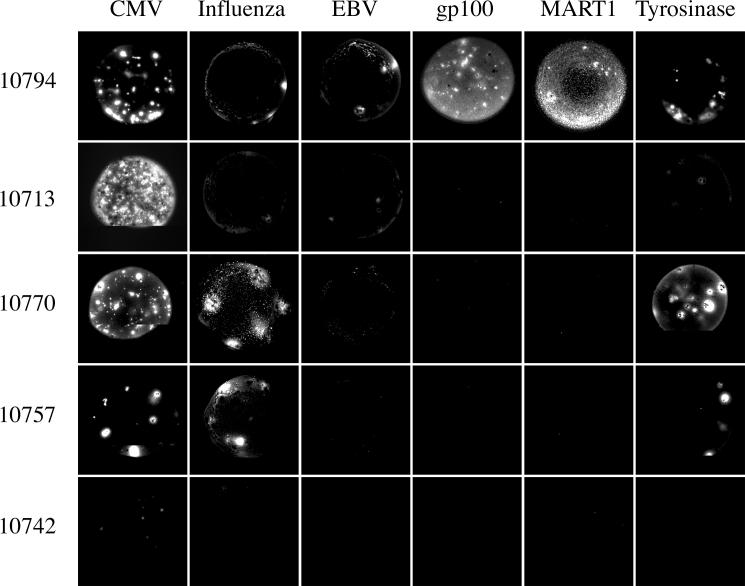
Methods and findings: In order to study the T cell responses in patients undergoing a melanoma-associated peptide vaccine trial, we have developed a high-throughput method using arrays of peptide-major histocompatibility complexes (pMHC) together with antibodies against secreted factors. T cells were specifically immobilized and activated by binding to particular pMHCs. The antibodies, spotted together with the pMHC, specifically capture cytokines secreted by the T cells. This technique allows rapid, simultaneous isolation and multiparametric functional characterization of antigen-specific T cells present in clinical samples. Analysis of CD8+ lymphocytes from ten melanoma patients after peptide vaccination revealed a diverse set of patient- and antigen-specific profiles of cytokine secretion, indicating surprising differences in their responsiveness. Four out of four patients who showed moderate or greater secretion of both interferon-gamma (IFNgamma) and tumor necrosis factor-alpha (TNFalpha) in response to a gp100 antigen remained free of melanoma recurrence, whereas only two of six patients who showed discordant secretion of IFNgamma and TNFalpha did so.

Conclusion: Such multiparametric analysis of T cell antigen specificity and function provides a valuable tool with which to dissect the molecular underpinnings of immune responsiveness and how this information correlates with clinical outcome.


Conflict of interest statement

Competing Interests: The authors have declared that no competing interests exist.

Figures

Figure 1
Figure 1. Peptide-MHC Cellular Microarrays
(A) A functional pMHC microarray diagram illustrating the array format. An array of spots containing different capture probes (pMHC molecules) cospotted with detector probes, which are antibodies against potentially secreted factors. Antigen-specific T cells are captured by recognition of printed pMHC, and may become activated. Subsequent secretion of specific factors is captured by the printed detector probes. The presence of those factors is detected by labeled secondary antibodies (developer probes). (B) Specificity of pMHC T cell capture is peptide-specific. Human CD8+ lymphocyte clones 132.2 and 461.30 were incubated on duplicate microarrays, which included gp100-2M/HLA-A2.1 and MART-1 M26/HLA-A2.1 tetramer and dimer spots. 2M-specific 132.2 cells were exclusively captured by gp100 spots, and M26-specific 461.30 binding was restricted to MART-1 spots. Binding efficiency to pMHC tetramer or dimer of a given specificity was similar.
Figure 2
Figure 2. Sensitivity of pMHC-Specific T-Cell Detection
The sensitivity of array-based T cell detection was compared to FACS-based detection. Patient PBMCs post-gp100 peptide vaccination were serially diluted in corresponding prevaccination sample. The dilution series was analyzed by both methods. Blue-labeled data correspond to the average number of bound cells per spot on the array (left graph) or the frequency of cells measured by FACS (right graph). The limit of detection in both methods was similar. Average number of bound cells per spot was obtained from five replicate spots on the same array. Error bars denote standard error of the mean across replicates. Red curves denote predicted results based on the values measured for the undiluted sample.
Figure 3
Figure 3. Profiling T Cell Function
Clonally derived MART-1/A2 specific human CD8+ T cells were incubated on a functional pMHC microarray on which individual spots contained pMHC or a control anti-CD8 monoclonal antibody (i.e., capture probes) cospotted with a panel of antibodies against potentially secreted factors (i.e., detector probes). MART-1-specific cells were immobilized on both MART-1/HLA-A2.1 and anti-CD8 containing spots. Bound cells were further incubated at 37 °C for 2 h. Secreted factors were captured by the coprinted antibodies at close vicinity to the secreting cells and detected using matched, biotinylated antibodies. Some of the initially bound cells detached during the staining procedure. (A) Top and bottom rows display IFNγ and TNFα secretions, respectively, each detected at single-cell resolution. The fluorescence images (red) are overlaid onto the differential interference contrast light microscopy images in the rightmost two columns. Not all immobilized T cells secreted detectable factors. No T cell binding or fluorescence was detectable on irrelevant pMHC spots (unpublished data). (B) Secretion profile for 17 different factors. Capture probes are either anti-CD8 antibody (left) or MART1 M26/A2 (right). Cospotted detector probes are indicated for each spot. Secretion signal is shown in pseudocolor, representing fluorescence intensity. Secreted factor-specific scaling has been applied to maximize resolution.
Figure 4
Figure 4. Anatomy of Cytokine Secretion
Secreted cytokine captured as it is released from activated lymphocytes immobilized on a pMHC cellular microarray shows cytokine-specific configurations. Select representative patient samples are shown for each labeled cytokine to illustrate the patterns of secretion for each individual cytokine.
Figure 5
Figure 5. Heterogeneity of Melanoma-Associated Antigen-Specific T Cell Responses following Peptide Vaccination
Eleven samples taken from patients enrolled in peptide vaccine trials were analyzed on pMHC functional microarrays. Patients received eight subcutaneous injections of peptides gp100 209–2M, MART1 M26, and tyrosinase 370D, along with adjuvant in a 6-mo period. Leukopheresis samples were collected after the eighth injection. Sample 10794 was collected from the same patient as 10735 after month 12. Functional profiles were generated by incubating patient CD8+ T lymphocytes on pMHC functional microarrays for 24 h at 37 °C and detecting the secreted factors with biotinylated secondary antibodies and streptavidin-phycoerythrin. Data were analyzed by automated fluorescence microscopy. Responses were scored on a five-point scale (0 to 4 bars), reflecting number of responders and overall fluorescent signal intensity per spot (Figure S1). Four bars indicate a strong response, and “0” indicates lack of a response. Each spot was printed in triplicate and analyzed individually. Patient clinical data are listed, including age and sex ( “ID”), stage of disease at enrollment (“Stage”), and outcome at follow-up (“Outcome”). Column labeled “IL12” specifies IL-12 adjuvant doses. Patient 10713 also received GM-CSF in addition to IL-12 adjuvant. Other secreted factors not shown include IL-4, IL-5, IL-10, IL-12p70, IL-1b, IL-3, IL-7, IL-13, IL-15, IL-17, lymphotactin, IP-10/CXCL10, TNFβ, VEGF, VEGF-D and granzyme A due to either lack of detectable secretion or limited analysis performed on only a fraction of the samples. In vitro restimulated cell lines directed against gp100 209 (132.2), MART1 M27 (461.30), or CMV pp65 495 (CMV94.3) were bound and secreted factors in response to gp100, MART1, and CMV (unpublished data), respectively.
Figure 6
Figure 6. Differences in Functional Profiles between Three Patients with Different Clinical Outcomes
gp100 209–2M spots co-spotted with anti-IFNγ, anti-TNFα, anti-IL1b, and anti-IL-6 are shown for three separate patient samples. Patient 10735, who remains free of disease at this writing, displays strong IFNγ, TNFα, IL-1b, and IL-6 secretion. Patients 10710 and 10737 show strong IFNγ secretion, but weak to no TNFα, IL-1b, and IL-6 secretion; both patients experienced disease recurrence soon after these samples were drawn. Note the diffuse pattern of IL-1b and IL-6 capture, which differs from the focal capture of IFNγ and TNFα.
Figure 7
Figure 7. Antigen-Specific Profiles within Individual Patients
CD8+ lymphocytes were isolated from PBMCs from patients 10794, 10713, 10770, 10757, and 10742, and incubated on a pMHC functional cellular microarray containing anti-IFNγ co-spotted with several different peptide-MHC (HLA-A2) complexes. These included melanoma-associated antigens gp100, MART-1, and tyrosinase, and viral antigens from cytomegalovirus, influenza virus, and Epstein-Barr virus (gp100 209–2M, MART1 M26, tyrosinase 370D, CMV pp65, influenza MP, and EBV BLF, respectively). The resulting IFNγ responses varied by antigen-specific T cell population.
Figure 8
Figure 8. Two Models of T-Cell Function
Acquisition and decay models depict two possible mechanisms that account for variability in the factors secreted by activated CD8+ T lymphocytes in response to antigen recognition. Acquisition refers to independent, sequential increases in responsiveness to activation, triggered by both cellular and secreted signals. Decay accounts for maximally functional T cells immediately upon completion of priming, after which signals, or time, lead to diminished responsiveness to activation.

Comment in

Similar articles

Cited by

References

    1. Rosenberg SA. Progress in human tumour immunology and immunotherapy. Nature. 2001;411:380–384. - PubMed
    1. van der Bruggen P, Traversari C, Chomez P, Lurquin C, De Plaen E, et al. A gene encoding an antigen recognized by cytolytic T lymphocytes on a human melanoma. Science. 1991;254:1643–1647. - PubMed
    1. Lee PP, Yee C, Savage PA, Fong L, Brockstedt D, et al. Characterization of circulating T cells specific for tumor-associated antigens in melanoma patients. Nat Med. 1999;5:677–685. - PubMed
    1. Talebi T, Weber JS. Peptide vaccine trials for melanoma: Preclinical background and clinical results. Semin Cancer Biol. 2003;13:431–438. - PubMed
    1. Kessels HW, Wolkers MC, Schumacher TN. Adoptive transfer of T-cell immunity. Trends Immunol. 2002;23:264–269. - PubMed

Publication types