. 2006 Jan 1;393(Pt 1):59-67.
doi: 10.1042/BJ20050674.

Endocytic processing of connexin43 gap junctions: a morphological study

Affiliations

Endocytic processing of connexin43 gap junctions: a morphological study

Edward Leithe et al. Biochem J. .

Abstract

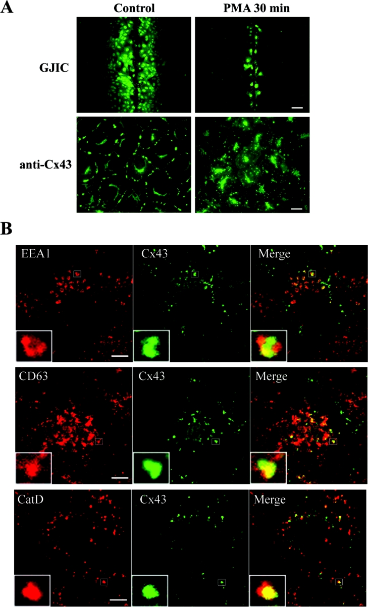
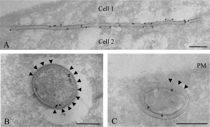
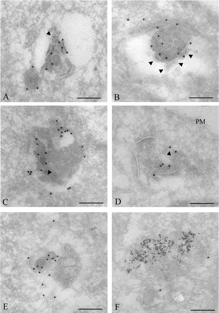
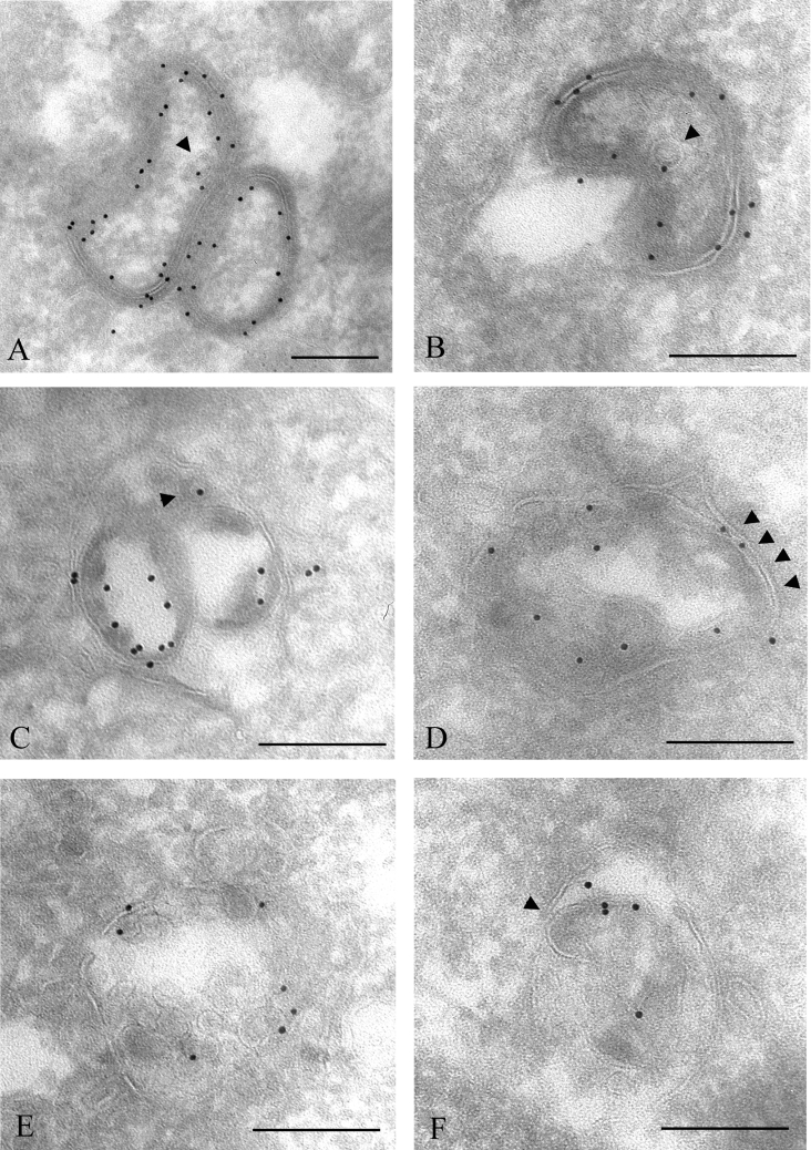
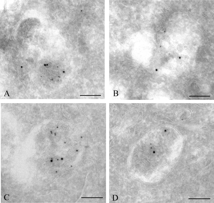
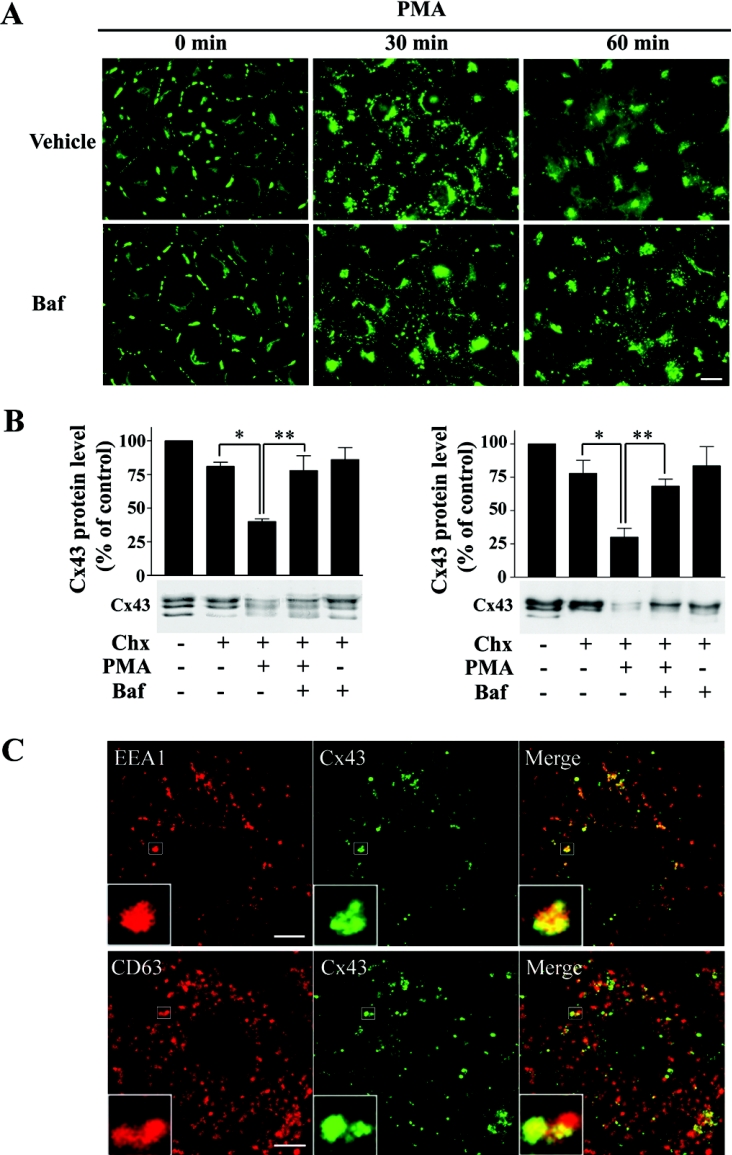
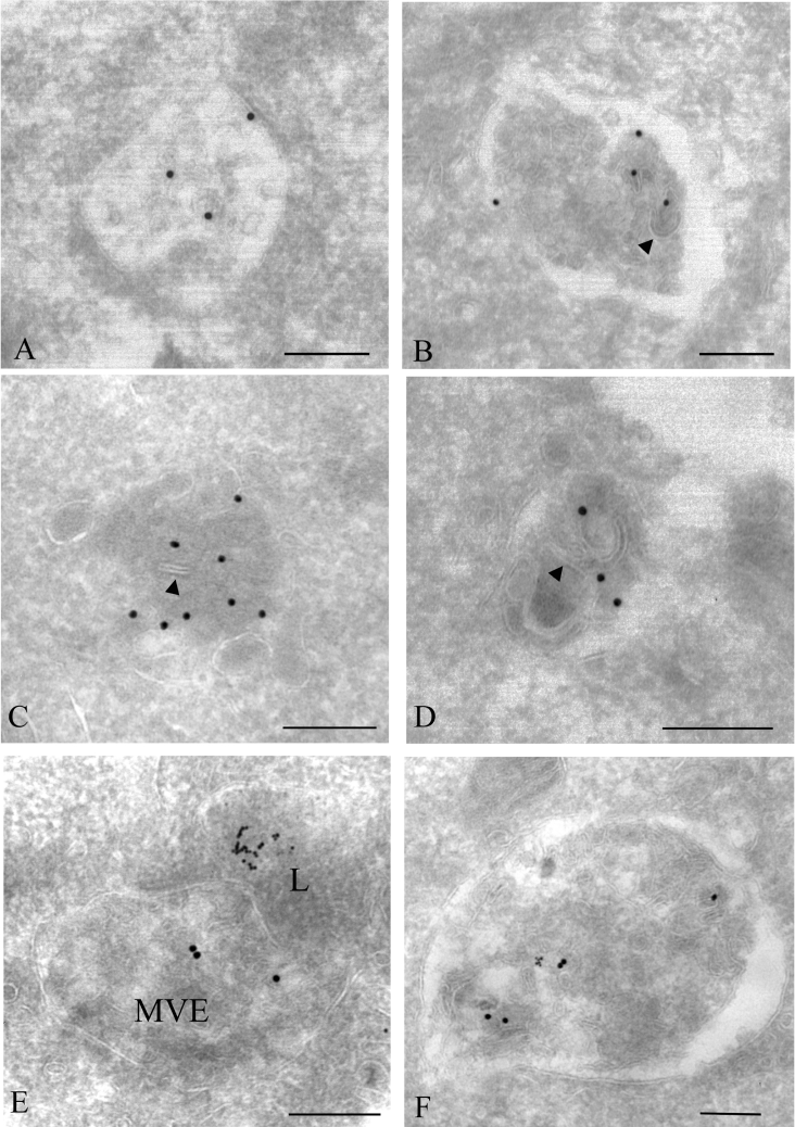
Gap junctions are plasma membrane areas enriched in channels that provide direct intercellular communication. Gap junctions have a high turnover rate; however, the mechanisms by which gap junctions are degraded are incompletely understood. In the present study, we show that in response to phorbol ester treatment, the gap junction channel protein Cx43 (connexin43) is redistributed from the plasma membrane to intracellular vesicles positive for markers for early and late endosomes and for the endolysosomal protease cathepsin D. Immunoelectron microscopy studies indicate that the double membranes of internalized gap junctions undergo separation and cutting, resulting in multivesicular endosomes enriched in Cx43 protein. Using preloading of BSA-gold conjugates to mark lysosomes, we provide evidence suggesting that the degradation process of the double-membrane structure of annular gap junctions occurs prior to transport of Cx43 to the lysosome. The results further suggest that bafilomycin A1, an inhibitor of vacuolar H+-ATPases, causes accumulation of Cx43 in early endosomes. Taken together, these findings indicate that internalized gap junctions undergo a maturation process from tightly sealed double-membrane vacuoles to connexin-enriched multivesicular endosomes with a single limiting membrane. The results further suggest that along with the processing of the double-membrane structure of annular gap junctions, connexins are trafficked via early and late endosomes, finally resulting in their endolysosomal degradation.


Figures

Figure 1
Figure 1. Immunofluorescence analysis of PMA-induced endocytosis of Cx43
(A) IAR20 cells were left untreated or treated with PMA (100 ng/ml) for 30 min. Gap junctional intercellular communication (GJIC) was examined by scrape loading of cells with Lucifer Yellow (upper panels; scale bar, 100 μm). Control IAR20 cells have extensive dye coupling, whereas PMA causes a nearly complete inhibition of dye transfer between cells. Cells were fixed, immunostained with anti-Cx43 antibodies and visualized using immunofluorescence microscopy (lower panels; scale bar, 20 μm). PMA causes redistribution of Cx43 from the plasma membrane to intracellular vesicles. (B) Cells were treated with PMA (100 ng/ml) for 30 min. Cells were fixed, double-stained with Cx43 antibody (middle panels) and EEA1, CD63 or cathepsin D antibody (left panels) as indicated and then visualized using confocal immunofluorescence microscopy. Merged images are shown in the right panels, with yellow indicating co-localization. Insets show enlarged view of representative Cx43-positive endosomes. Scale bars, 10 μm.
Figure 2
Figure 2. Immunoelectron microscopy of intercellular and annular Cx43 gap junctions
To identify lysosomes, cells were incubated with 8 nm BSA–gold for 3 h and chased overnight. Cells were left untreated or treated with PMA (100 ng/ml) for 30 min and fixed. Cryosections of untreated cells (A) or cells treated with PMA (100 ng/ml) for 30 min (B, C) were labelled for Cx43 (15 nm gold). (A) In untreated cells, Cx43 gap junction plaques were frequently observed between adjacent cells. (B, C) Annular gap junctions. Large areas of the gap junction double membranes appear to have started to separate (arrowheads). PM, plasma membrane. Scale bars, 200 nm.
Figure 3
Figure 3. Immunoelectron microscopy analysis of PMA-induced Cx43 gap junction degradation
To identify lysosomes, cells were incubated with 8 nm BSA–gold for 3 h and chased overnight. Cells were treated with PMA (100 ng/ml) for 30 min, fixed, prepared for electron microscopy and labelled for Cx43 (15 nm gold). (A) An annular gap junction appears to have started to be cut into smaller membrane pieces. Some parts of the gap junction membrane appear to form a small intravacuolar vesicle (arrowhead). (B) The double membrane of the annular gap junction appears to be cut and to separate. The resulting single membranes appear to form small vesicles (arrowheads), similar to vesicles inside multivesicular endosomes. (CE) Cx43 labelling in multivesicular endosomes. Some of the intravacuolar vesicles have irregular shapes and are different from vesicles usually seen in multivesicular endosomes. Some membrane areas of the endosomes appear to be remnants of double-membrane gap junctions (arrowheads). (F) Cx43 labelling in a lysosome, identified as an electron-dense vesicle containing endocytosed 8 nm gold particles. PM, plasma membrane. Scale bars, 200 nm.
Figure 4
Figure 4. Immunoelectron microscopy analysis of EGF-induced Cx43 gap junction degradation
IAR20 cells were treated with EGF (50 ng/ml) for 30 min. The cells were fixed, prepared for electron microscopy and labelled for Cx43 (10 nm gold). (A) Two tightly apposed annular gap junctions. Some parts of the double membranes appear to have started to separate. One single-membrane vesicle inside the annular gap junction is labelled with one gold particle (arrowhead). (B) An annular gap junction appears to fuse with an endosome. The annular gap junction appears to invaginate into itself. One single-membrane vesicle inside the annular gap junction is labelled with one gold particle (arrowhead). (C) An annular gap junction containing two large and one small (arrowhead) internal vesicles with Cx43 labelling. At some places, the vesicles appear to be tightly sealed with the outer membrane, similar to what is observed between the two membranes of gap junctions. (DF) Cx43 is localized in multivesicular endosomes. Some of the intravacuolar vesicles have irregular shapes and are different from vesicles usually seen in multivesicular endosomes. Some of the vesicles are tightly associated with the outer membrane, forming double membranes that appear to be remnants of gap junctions (arrowheads). Scale bars, 200 nm.
Figure 5
Figure 5. Immunoelectron localization of Cx43 in early and late endosomes
(A, B) IAR20 cells were treated with PMA (100 ng/ml) for 30 min. During the last 10 min of incubation, 8 nm BSA–gold particles were added to the medium, to mark early endosomes. Cells were fixed, prepared for electron microscopy and labelled for Cx43 (15 nm gold). Early endosomes, characterized by their electron-lucent appearance, a few internal vesicles and content of 8 nm gold particles, were often found to contain Cx43. Cx43 was found both on the limiting vacuolar membrane and on intravacuolar vesicles. (C, D) To mark both early and late endosomes, cells were treated with PMA (100 ng/ml) for 30 min in a medium containing 8 nm BSA–gold particles. Cells were fixed and labelled for Cx43 (15 nm gold). Cx43 was found in late endosomes, differentiated from early endosomes by their globular form, their more electron-dense appearance and by containing more internal vesicles. Scale bars, 200 nm.
Figure 6
Figure 6. Effect of bafilomycin A1 on PMA-induced endocytosis of Cx43
(A) IAR20 cells were treated with bafilomycin A1 (Baf; 200 nM) or vehicle (DMSO) for 30 min prior to and during treatment with PMA (100 ng/ml) for the indicated times. Cells were fixed, immunostained with anti-Cx43 antibodies and visualized using fluorescence microscopy. Scale bar, 20 μm. (B) IAR20 cells were left untreated or treated with bafilomycin A1 (200 nM) or vehicle (DMSO) for 30 min prior to and during treatment with PMA (100 ng/ml) and cycloheximide (Chx; 10 μg/ml) or cycloheximide alone for 60 min as indicated. Cell lysates were prepared and equal amounts of total cell protein were subjected to SDS/PAGE. Cx43 was detected by Western blotting using antibodies raised against the C-terminal end of Cx43 (left panel) or antibodies recognizing the amino acids 130–143 in the internal loop of Cx43 (Sigma antibody; right panel). For both antibodies, the Cx43 band intensities on gels were measured using Scion Image. Results shown are the means±S.E.M. for three independent experiments. Statistical analysis was performed using one-way ANOVA with Tukey–Kramer multiple comparisons test. Cells treated with cycloheximide and PMA had significantly reduced levels of Cx43 protein compared with cells treated with cycloheximide alone (*P<0.05). This loss of Cx43 was significantly counteracted by bafilomycin A1 (**P<0.05). (C) IAR20 cells were treated with bafilomycin A1 (200 nM) for 30 min prior to and during treatment with PMA (100 ng/ml) for 60 min. Cells were fixed and double stained with Cx43 (middle panels) and EEA1 or CD63 (left panels) antibodies as indicated and visualized using confocal immunofluorescence microscopy. Merged images are shown in the right panels, with yellow indicating co-localization. Insets show enlarged view of representative Cx43-positive endosomes. Scale bars, 10 μm.
Figure 7
Figure 7. Immunoelectron microscopy analysis of Cx43 localization in cells treated with bafilomycin A1 and PMA
To identify lysosomes, cells were incubated with 8 nm BSA–gold for 3 h and chased overnight. IAR20 cells were treated with bafilomycin A1 (200 nM) for 30 min prior to and during treatment with PMA (100 ng/ml) for 60 min, fixed, prepared for electron microscopy and labelled for Cx43 (15 nm gold). (A) Cx43 often localized in electron-lucent vacuoles with few internal vesicles, which resembled early endosomes. Cx43 was, under these conditions, found both on the limiting membrane of the vacuole and on the internal vesicles. (BD) Cx43 was also found in more electron-lucent vacuoles containing several internal vesicles. Sometimes these vacuoles contained what appeared to be remnants of gap junction double membranes (arrowheads). (E) Cx43 labelling in multivesicular endosome that appear to fuse with a lysosome. (F) Cx43 labelling in enlarged multivesicular endosome with 8 nm gold particles. Scale bars, 200 nm.

References

    1. Saez J. C., Berthoud V. M., Branes M. C., Martinez A. D., Beyer E. C. Plasma membrane channels formed by connexins: their regulation and functions. Physiol. Rev. 2003;83:1359–1400. - PubMed
    1. Musil L. S., Goodenough D. A. Multisubunit assembly of an integral plasma membrane channel protein, gap junction connexin43, occurs after exit from the ER. Cell (Cambridge, Mass.) 1993;74:1065–1077. - PubMed
    1. Sosinsky G. E. Molecular organization of gap junction membrane channels. J. Bioenerg. Biomembr. 1996;28:297–309. - PubMed
    1. Segretain D., Falk M. M. Regulation of connexin biosynthesis, assembly, gap junction formation, and removal. Biochim. Biophys. Acta. 2004;1662:3–21. - PubMed
    1. Wei C. J., Xu X., Lo C. W. Connexins and cell signaling in development and disease. Annu. Rev. Cell Dev. Biol. 2004;20:811–838. - PubMed

Publication types

LinkOut - more resources