Review
. 2005 Oct;18(5):322-36.
doi: 10.1111/j.1600-0749.2005.00269.x.

The Silver locus product Pmel17/gp100/Silv/ME20: controversial in name and in function

Affiliations
Review

The Silver locus product Pmel17/gp100/Silv/ME20: controversial in name and in function

Alexander C Theos et al. Pigment Cell Res. 2005 Oct.

Abstract

Mouse coat color mutants have led to the identification of more than 120 genes that encode proteins involved in all aspects of pigmentation, from the regulation of melanocyte development and differentiation to the transcriptional activation of pigment genes, from the enzymatic formation of pigment to the control of melanosome biogenesis and movement [Bennett and Lamoreux (2003) Pigment Cell Res. 16, 333]. One of the more perplexing of the identified mouse pigment genes is encoded at the Silver locus, first identified by Dunn and Thigpen [(1930) J. Heredity 21, 495] as responsible for a recessive coat color dilution that worsened with age on black backgrounds. The product of the Silver gene has since been discovered numerous times in different contexts, including the initial search for the tyrosinase gene, the characterization of major melanosome constituents in various species, and the identification of tumor-associated antigens from melanoma patients. Each discoverer provided a distinct name: Pmel17, gp100, gp95, gp85, ME20, RPE1, SILV and MMP115 among others. Although all its functions are unlikely to have yet been fully described, the protein clearly plays a central role in the biogenesis of the early stages of the pigment organelle, the melanosome, in birds, and mammals. As such, we will refer to the protein in this review simply as pre-melanosomal protein (Pmel). This review will summarize the structural and functional aspects of Pmel and its role in melanosome biogenesis.


Figures

Figure 1
Figure 1
Human pre-melanosomal protein (Pmel) gene and splice isoforms. (A) Organization of human SILV (Pmel) gene, according to Bailin et al. (1996). Exons are indicated by boxes, and introns by lines, drawn to scale (except where sizes of introns are indicated). The numbers at bottom indicate the codons at the start of each exon; 5′- and 3′-untranslated regions (5′UT, 3′UT) are indicated in gray. Hatched regions indicate alternatively spliced exons. (B) Schematic diagram of the products of the four known human Pmel splice isoforms. The intermediate (int.) form is the most abundant, and the short forms (short-l and short-i, including or lacking, respectively, the sequence uniquely present in the long but not the intermediate form), are the least abundant. ss, signal sequence; tm, transmembrane domain; cyt., cytoplasmic domain. Numbers indicate the amino acids at the borders of the topologic domains (ss, lumenal, tm and cyt.) or of the alternatively spliced regions with respect to Pmel-long.
Figure 2
Figure 2
Domains within pre-melanosomal protein (Pmel) and their identity among vertebrates. (A) Individual domains within the human Pmel long form (identified in the text) are shown in different colors, with amino acid positions for start and end of each domain indicated below. Consensus sites for addition of N-linked oligosaccharides are indicated by branched structures; a site near the beginning of the repeat (RPT) domain is noted in gray because it is not conserved and unlikely used (Maresh et al., 1994b and our unpublished observations). Circles indicate positions of cysteine residues. TM, transmembrane domain; Cyt., cytoplasmic domain; CS, cleavage site. (B) Amino acid sequence comparison of Pmel orthologs in indicated species. The GenBank accession numbers from which the sequences were taken are indicated in the text; only the long form of human Pmel is shown. Red text indicates amino acid identities among all sequenced orthologs; orange indicates amino acid homology among most orthologs. Individual domains are indicated by bars over the sequence colored as in (A) and (C); the domain definitions are extended beyond the borders in (A) to cover the entire sequence, and were used in the analysis described in (C). (C) The amino acid sequence of each of the indicated domains of human Pmel was compared with that of the corresponding domain in each of the indicated species. The identity of the corresponding amino acid sequence to that of human Pmel is indicated graphically for each domain. Domain colors correspond to those in panel (A); also included in the comparison is the sequence of human Nmb.
Figure 3
Figure 3
Melanosome stages of development. (A) Schematic diagram of the four melanosome stages as described in the text. (B) Representative electron micrograph of an ultrathin section of plastic-embedded MNT-1 cells showing examples of each of the melanosome stages I, II, III and IV as indicated. Lys, lysosome; m, mitochondria.
Figure 4
Figure 4
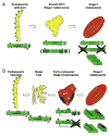
Two models for pre-melanosomal protein (Pmel) trafficking to stage I/II melanosomes. Shown are two models for Pmel trafficking through the cell, with schematic diagram of the processing intermediate for Pmel within each organelle as described in the text. (A) Based on models described in Kushimoto et al. (2001) and Yasumoto et al. (2004). (B) Based on models described in Berson et al. (2001, and Raposo et al. (2001). The Pmel lumenal, transmembrane (TM; white) and cytoplasmic (cyt) domains are labeled, and the RPT domain is indicated by vertical shading. N-glycosylation sites are indicated by branched structures, and potential O-glycosylation sites are indicated by circles; branching in panel (B) indicates processing of glycans in the Golgi. Cleavage products that are eventually lost to proteolytic degradation are indicated in dark green, and oligomerization of the final cleavage products is indicated by stacking. Note that the cleavage boundaries and responsible proteases for generating the final products (HMB-45-reactive RPT region and HMB-50/NKI-beteb-reactive N-terminal region) in stage II melanosomes have not yet been defined in either model.
Figure 5
Figure 5
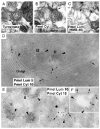
Immunoelectron microscopy evidence for pre-melanosomal protein (Pmel) cytoplasmic domain in melanosome precursors. Published electron micrographs of immunogold labeled samples derived from the pigmented MNT-1 melanoma cell line. Immunogold labeling for the Pmel cytoplasmic domain is indicated by arrows, and for the lumenal domain by arrowheads. (A–C) Pre-embedding immunogold labeling of purified stage II melanosome fractions using antibodies to the tyrosinase cytoplasmic domain [(A); stars], the Pmel cytoplasmic domain [(B), αPep13h; arrows] or the Pmel lumenal domain [(C), HMB-45; arrowheads]. Note the cluster of gold between stage II melanosomes in (B), contrasting with the even distribution of gold on the internal striations in (C) and on the limiting membrane of the melanosome in (A). Data are reproduced from Figure 4 of Kushimoto et al. (2001) (D–F). Double immunogold labeling of ultrathin cryosections of MNT-1 cells using antibodies to the Pmel cytoplasmic domain (arrows) or lumenal domain (HMB-50; arrowheads). (D). Reproduced from Supplementary Figure S3 of Raposo et al., 2001. Pmel cytoplasmic domain is labeled with αPep13h and protein A-conjugated 15-nm gold (arrows), and HMB-50 labeling is developed with protein A-conjugated 10-nm gold (arrowheads). Note the labeling of the Golgi stacks by both antibodies, whereas the stage II melanosomes are labeled only in the lumen by HMB-50. (E,F). Reproduced from Figure 6 of Berson et al. (2003). Pmel cytoplasmic domain is labeled with αPep13h and protein A-conjugated 10-nm gold (arrows), and HMB-50 labeling is developed with protein A-conjugated 5-nm gold (arrowheads). In (E), note the labeling of stage II melanosomes only by HMB-50, whereas an endosomal structure shows labeling for both antibodies. (F), a typical stage I melanosome, in which cytoplasmic domain labeling is evident at the limiting membrane.

Comment in

References

    1. Adema GJ, de Boer AJ, van't Hullenaar R, Denijn M, Ruiter DJ, Vogel AM, Figdor CG. Melanocyte lineage-specific antigens recognized by monoclonal antibodies NKI-beteb, HMB-50, and HMB-45 are encoded by a single cDNA. Am J Pathol. 1993;143:1579–1585. - PMC - PubMed
    1. Adema GJ, de Boer AJ, Vogel AM, Loenen WAM, Figdor CG. Molecular characterization of the melanocyte lineage-specific antigen gp100. J Biol Chem. 1994;269:20126–20133. - PubMed
    1. Badman MK, Shennan KI, Jermany JL, Docherty K, Clark A. Processing of pro-islet amyloid polypeptide (proIAPP) by the prohormone convertase PC2. FEBS Lett. 1996;378:227–231. - PubMed
    1. Bailin T, Lee ST, Spritz RA. Genomic organization and sequence of D12S53E (Pmel 17), the human homologue of the mouse silver (si) locus. J Invest Dermatol. 1996;106:24–27. - PubMed
    1. Bakker ABH, Schreurs MWJ, de Boer AJ, Kawakami Y, Rosenberg SA, Adema GJ, Figdor CG. Melanocyte lineage-specific antigen gp100 is recognized by melanoma-derived tumor-infiltrating lymphocytes. J Exp Med. 1994;179:1005–1009. - PMC - PubMed

Publication types

MeSH terms