Comparative Study
. 2005 Oct;18(5):349-59.
doi: 10.1111/j.1600-0749.2005.00249.x.

MITF and cell proliferation: the role of alternative splice forms

Affiliations
Comparative Study

MITF and cell proliferation: the role of alternative splice forms

Keren Bismuth et al. Pigment Cell Res. 2005 Oct.

Abstract

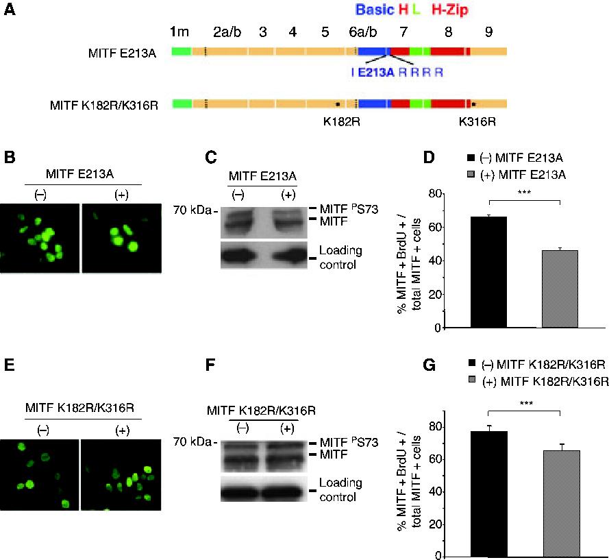
Recent studies show that the melanocyte transcription factor MITF not only activates differentiation genes but also genes involved in the regulation of the cell cycle, suggesting that it provides a link between cell proliferation and differentiation. MITF, however, comes in a variety of splice isoforms with potentially distinct biological activities. In particular, there are two isoforms, (-) and (+) MITF, that differ in six residues located upstream of the DNA binding basic domain and show slight differences in the efficiency with which they bind to target DNA. Using in vitro BrdU incorporation assays and FACS analysis in transiently transfected cells, we show that (+) MITF has a strong inhibitory effect on DNA synthesis while (-) MITF has none or only a mild one. The strong inhibitory activity of (+) MITF is not influenced by a number of mutations that modulate MITF's transcriptional activities and is independent of the protein's carboxyl terminus but dependent on its aminoterminus. A further dissection of the molecule points to the importance of an aminoterminal serine, serine-73, which in both isoforms is phosphorylated to comparable degrees. The results suggest that one or several aminoterminal domains cooperate with the alternatively spliced hexapeptide to render MITF anti-proliferative in a way that does not depend on direct E box binding.


Figures

Figure 1.
Figure 1.
(+) MITF but not (-) MITF inhibits BrdU incorporation in transfected cells. (A) MITF immunofluorescence of HEK293 cells transfected with (-) or (+) Mitf cDNA. (B) Western blot analysis of HEK293 cell extracts prepared 20 h after transfection with either (-) or (+) Mitf cDNA, using C5 anti-MITF antibody. Loading control corresponds to an unspecific band. (C) BrdU incorporation in HEK293 cells 20 h after transfection with (-) or (+) Mitf cDNA or pEGFP-C1 plasmid. Cells were incubated for 30 min with 10 μM BrdU and double-labeled with anti-BrdU and C5 anti-MITF antibodies. Double-positive cells were counted as a percentage of the total number of MITF-positive cells. (D) Similar assay as in C but done in BHK cells.
Figure 2.
Figure 2.
MITF mutations that affect DNA binding or transcriptional activity do not interfere with the enhanced inhibition of BrdU incorporation by (+) MITF. (A) schematic representation of MITF E213A and MITF K182R/316R. (B and E) Immunofluorescent labeling of cells transfected with (-) or (+) MITF E213A (B) or (-) or (+) MITF K182R/K316R (E). (C and F) Western blot analysis of cells transfected with (-) or (+) MITF E213A (C) or (-) or (+) MITF K182R/K316R (F). Loading controls correspond to beta-tubulin. (D and G) BrdU incorporation in cells transfected with (-) or (+) MITF E213A (D) or (-) or (+) MITF K182R/K316R (G). Cells were incubated with 10 μM BrdU and stained with anti-MITF and anti-BrdU. Double MITF/BrdU-positive cells were counted as in Figure 1.
Figure 3.
Figure 3.
A mutation leading to decreased nuclear translocation reduces the inhibitory effect of (+) MITF. (A) schematic representation of MITF DelR216. (B) Cytoplasmic and nuclear localization of (-) and (+) MITF DelR216. (C) Western blot analysis of protein extracts of cells transfected with (-) and (+) MITF DelR216. Loading control corresponds to beta-tubulin. (D) BrdU incorporation assay in cells transfected with (-) or (+) MITF DelR216. Double MITF/BrdU-positive cells were counted as in Figure 1.
Figure 4.
Figure 4.
Effects of truncated or serine-73-to-alanine mutated (-) and (+) MITF on BrdU incorporation. (A) Schematic representation of the different mutants. (B, E and H) MITF immunofluorescence of HEK293 cells transfected with the indicated (-) or (+) MITF mutants. (C, F and I) Western blot analyses of protein extracts from cells transfected with the indicated (-) or (+) MITF mutants. Loading control corresponds to beta-tubulin (C and F) or an unspecific band (I). (D, G and J) BrdU incoporation in cells transfected with the indicated (-) or (+) MITF mutants. BrdU incorporation and counting done as in Figure 1.
Figure 5.
Figure 5.
Cell cycle FACS analysis on HEK293 cells transfected with wild type (-) and (+) MITF WT. Twenty hours after transfection, cells were incubated with BrdU for 30 min and processed for BrdU and propidium iodide labeling before FACS analysis. (A) FACS plots of representative assays. Control represents non-transfected cells, and (-) MITF and (+) MITF correspond to transfections with the respective plasmids. The gates (R1-R10) were determined empirically. Note that HEK293 cells are aneuploid (hypotriploid) but undergo normal DNA doubling with each round of replication. R1, apoptotic, (hypo-)hypotriploid cells with no BrdU incorporated; and R2, apoptotic, (hypo)-hypotriploid cells that have incorporated BrdU. R6, apoptotic cells corresponding to (hypo-)hypohexaploidy. R3, G0G1; R5, G2M; R7, early S-phase; R8, late S-phase; R9, S-phase with low BrdU content; R10, S-phase with high BrdU content. Note that total S-phase corresponds to R9 + R10. (B-D) FACS analysis was repeated in three independent experiments, using the same gating as shown in (A). The results were quantified using the Cellquest software. (B) Quantitative analysis of the percentage of cells in G0G1, S, and G2M phases of the cell cycle. Note a significant difference between (-) and (+) MITF on S-phase (P < 0.02). (C) Separation of S-phase into low and high BrdU content. Note a significant difference between (-) and (+) MITF in low BrdU content (P < 0.01). (D) Separation of cells into early and late S-phase. Note a significant difference between (-) and (+) MITF on early S-phase (P < 0.01).

References

    1. Arnheiter H, Hou L, Nguyen M-TT, Bismuth K, Csermely T, Murakami H, Skuntz S, Liu W, Bharti K.Mitf - A Matter of Life and Death for the Developing Melanocytes Humana Press; Totowa, NJ: in press
    1. Carreira S, Liu B, Goding CR. The gene encoding the T-box factor Tbx2 is a target for the microphthalmia-associated transcription factor in melanocytes. J. Biol. Chem. 2000;275:21920–21927. - PubMed
    1. Carreira S, Goodall J, Aksan I, La Rocca SA, Galibert MD, Denat L, Larue L, Goding CR. Mitf cooperates with Rb1 and activates p21Cip1 expression to regulate cell cycle progression. Nature. 2005;433:764–769. - PubMed
    1. Crescenzi M, Fleming TP, Lassar AB, Weintraub H, Aaronson SA. MyoD induces growth arrest independent of differentiation in normal and transformed cells. Proc. Natl. Acad. Sci. U. S. A. 1990;87:8442–8446. - PMC - PubMed
    1. Del Bene F, Tessmar-Raible K, Wittbrodt J. Direct interaction of geminin and Six3 in eye development. Nature. 2004;427:745–749. - PubMed

Publication types

MeSH terms