Comparative Study
. 2005 Oct;18(5):370-81.
doi: 10.1111/j.1600-0749.2005.00263.x.

Different approaches for assaying melanosome transfer

Affiliations
Comparative Study

Different approaches for assaying melanosome transfer

Werner Berens et al. Pigment Cell Res. 2005 Oct.

Abstract

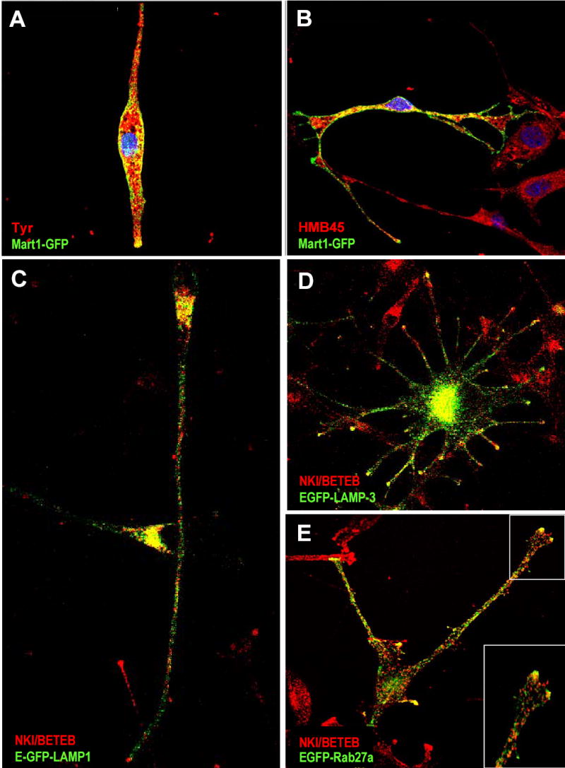
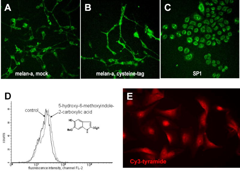
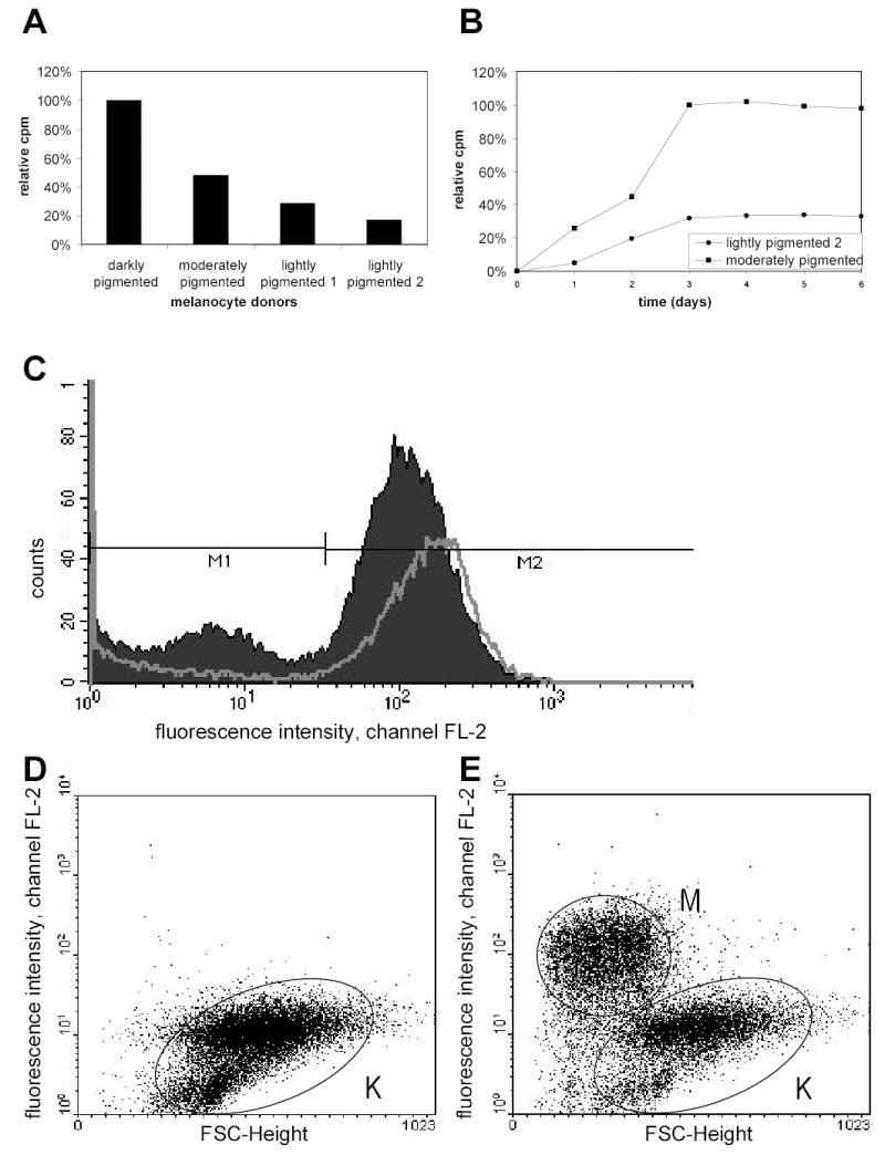
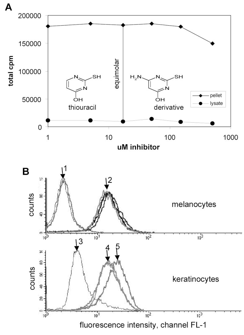
Many approaches have been tried to establish assays for melanosome transfer to keratinocytes. In this report, we describe and summarize various novel attempts to label melanosomes in search of a reliable, specific, reproducible and quantitative assay system. We tried to fluorescently label melanosomes by transfection of GFP-labeled melanosomal proteins and by incubation of melanocytes with fluorescent melanin intermediates or homologues. In most cases a weak cytoplasmic fluorescence was perceived, which was probably because of incorrect sorting or deficient incorporation of the fluorescent protein and different localization. We were able to label melanosomes via incorporation of 14C-thiouracil into melanin. Consequently, we tried to develop an assay to separate keratinocytes with transferred radioactivity from melanocytes after co-culture. Differential trypsinization and different magnetic bead separation techniques were tested with unsatisfactory results. An attempt was also made to incorporate fluorescent thiouracil, since this would allow cells to be separated by FACS. In conclusion, different methods to measure pigment transfer between donor melanocytes and acceptor keratinocytes were thoroughly examined. This information could give other researchers a head start in the search for a melanosome transfer assay with said qualities to better understand pigment transfer.


Figures

Figure 1
Figure 1. Transfection of human melanocytes with E(C)GFP-CYT-TYR
A) schematic representation of the GFP constructs of TYR and TYRP1, showing the position of the fluorescent moiety. B) Human melanocytes (culture method 2) transfected with E(C)GFP-CYT-TYR demonstrate diffuse green fluorescence with perinuclear accentuation. D) Addition of bafilomycin A1 (25 nM) results in a brighter signal that shows a clear granular distribution. F) Co-staining with the melanosomal marker NKI/BETEB (against GP100) demonstrates discrete co-localisation in the perinuclear region but not in the dendrite tips.
Figure 2
Figure 2. Transfection of human and mouse melanocytes with melanosome-specific and -nonspecific GFP constructs
A) Co-staining of melan-a melanocytes transfected with Mart1-GFP and α-PEP7 (against Tyr). B) Co-staining of Mart1-GFP transfected melan-a melanocytes with HMB45 (against Gp100). C) Human melanocytes (culture method 2) transfected with LAMP1-GFP were stained with NKI/BETEB (against GP100); co-localization is seen in the perinuclear area, but not in the dendrites. Co-staining with α-PEP1 (against TYRP1) gives the same result (not shown). D) Human melanocytes transfected with LAMP3-GFP were stained with NKI/BETEB; co-localisation in the dendrites is discrete. E) GFP fluorescence of EGFP-RAB27a transfected human melanocytes and co-staining with NKI/BETEB (against GP100) co-localize at the cell periphery, especially at the dendrite tips.
Figure 3
Figure 3. Cysteine-tag methods to fluorescently label melanosomes and incubation with Cy3-tyramide
A-B) Melan-a melanocytes were transfected with mock- (A) or cysteine-tag DNA (B); staining was done with 2 μM FlAsH and 10 μM EDT 2 days after transfection. (C) SP1 keratinocytes stained as in (B). D) Melan-a melanocytes were incubated for 3 days with 5,6-dihydroxy-indole- 2-carboxylic acid and were analysed by FACS; a slight fluorescence is detected. E) Human melanocytes (culture method 2) incubated with Cy3-tyramide (25nM-5μM) for 4 h display a reticular fluorescent pattern with perinuclear accentuation.
Figure 4
Figure 4. Identification of melanocytes by 14C-thiouracil and by phycoerythrin labeled antibodies
A) Primary human melanocytes with different degrees of pigmentation were incubated with 1 μCi 14C-thiouracil for 3 days; data reported show relative uptake of label. B) Two different human melanocyte monocultures were incubated with 1 μCi 14C-thiouracil for 3 days, the medium was replaced, and incorporation was measured for another 3 days. C) Keratinocytes and melanocytes in monoculture were stained with PE-labeled CD49f against keratinocyte-specific α-6 integrin, mixed and incubated with anti-PE beads from Miltenyi. Intervals M1 for melanocytes and M2 for keratinocytes were analyzed for values before and after magnetic separation. D–E) Keratinocytes and melanocytes in co-culture were stained with CD117-PE against melanocyte-specific c-kit, and incubated with anti-PE beads from Miltenyi. The negative fraction exclusively contained unlabeled cells, representing keratinocytes (K) (D). An important fraction of keratinocytes was present in the positive fraction in addition to the selected melanocytes (M) (E). (A–C, culture method 1; D–E, culture method 2).
Figure 5
Figure 5. Competition of 14C-thiouracil incorporation into melanin by a thiouracil derivative and CFDA labeling of melanocytes
A) Human melanocytes were incubated with 1 μM 14C-thiouracil (17 μM) and different concentrations of 4-amino-6-hydroxy-2-mercaptopyrimidine for 3 days and the 14C content was measured in cell pellets and lysates by liquid scintillation counting. B) Fluorescence of human primary melanocytes and keratinocytes with or without CFDA labeling for 30 min and PE-αCD49f stain for keratinocytes after 3 days of incubation. Samples were incubated with pigmentation modifiers (10 μM forskolin or 150 μM α-lipoic acid). Upper graph: Arrow 1 shows 2 unstained melanocyte samples, arrow 2 from left to right the CFDA treated samples in combination with 10 μM forskolin-(light grey curve), 0.02% DMSO-treated control (black) and 30 μM lipoic acid (dark grey).. Lower graph: Control keratinocytes without CFDA labeling, arrow 3 shows an unstained sample, arrow 4 and 5 PE-αCD49f stained samples, with arrow 4 showing a 0.02% DMSO treated control (light grey) and a 30 μM lipoic acid (dark grey) treated sample, and arrow 5 a 10 μM forskolin treated sample. (culture method 1)

References

    1. Adams SR, Campbell RE, Gross LA, Martin BR, Walkup GK, Yao Y, Llopis J, Tsien RY. New biarsenical ligands and tetracysteine motifs for protein labeling in vitro and in vivo: synthesis and biological applications. J Am Chem Soc. 2002;124:6063–6076. - PubMed
    1. Ancans J, Thody AJ. Activation of melanogenesis by vacuolar type H(+)-ATPase inhibitors in amelanotic, tyrosinase positive human and mouse melanoma cells. FEBS Lett. 2000;478:57–60. - PubMed
    1. Babiarz-Magee L, Chen N, Seiberg M, Lin CB. The expression and activation of protease-activated receptor-2 correlate with skin color. Pigment Cell Res. 2004;17:241–251. - PubMed
    1. Bahadoran P, Busca R, Chiaverini C, Westbroek W, Lambert J, Bille K, Valony G, Fukuda M, Naeyaert JM, Ortonne JP, Ballotti R. Characterization of the molecular defects in Rab27a, caused by RAB27A missense mutations found in patients with Griscelli syndrome. J Biol Chem. 2003;278:11386–11392. - PubMed
    1. Basrur V, Yang F, Kushimoto T, Higashimoto Y, Yasumoto K, Valencia J, Muller J, Vieira WD, Watabe H, Shabanowitz J, et al. Proteomic analysis of early melanosomes: identification of novel melanosomal proteins. J Proteome Res 2003;2:69–79. - PubMed

Publication types

MeSH terms