. 2005 Oct;116(2):276-86.
doi: 10.1111/j.1365-2567.2005.02222.x.

Tyrosine kinase activity and remodelling of the actin cytoskeleton are co-temporally required for degranulation by cytotoxic T lymphocytes

Affiliations

Tyrosine kinase activity and remodelling of the actin cytoskeleton are co-temporally required for degranulation by cytotoxic T lymphocytes

Aimee Shen et al. Immunology. 2005 Oct.

Abstract

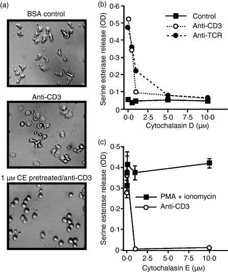
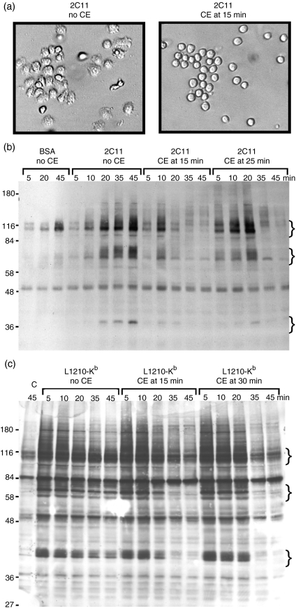
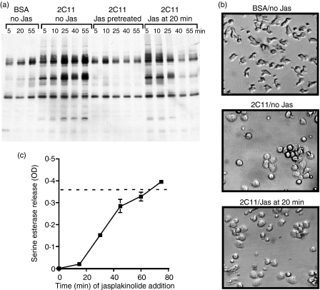
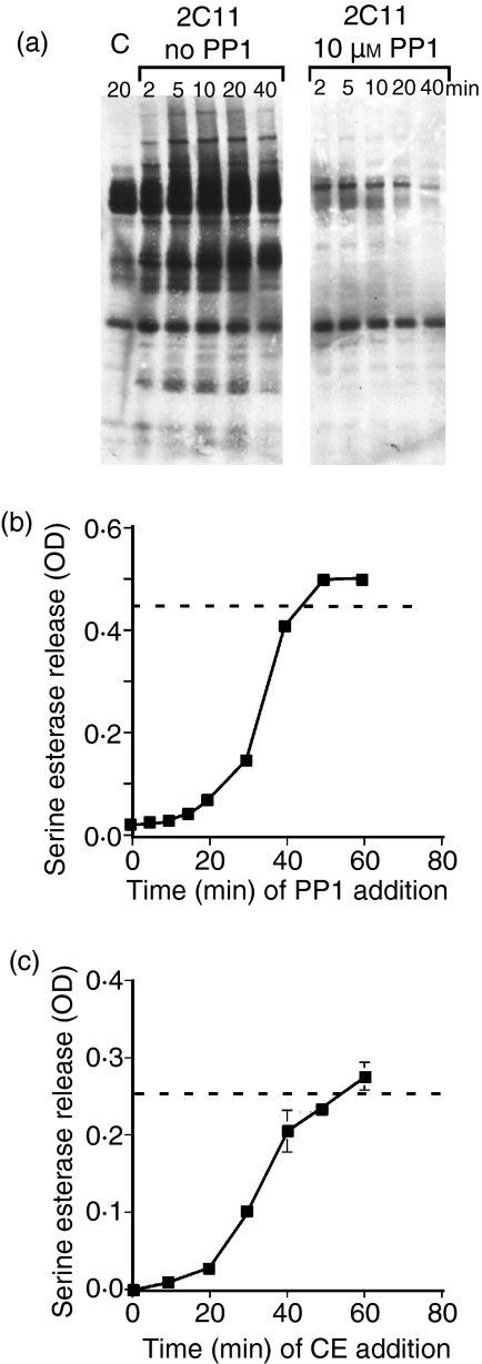
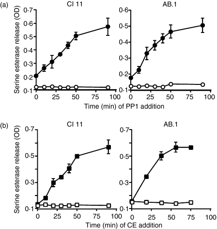
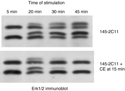
In this study, we examined the contribution of the actin cytoskeleton to T-cell receptor (TCR)-initiated signalling in cytotoxic T lymphocytes (CTLs). We demonstrate that cytoskeletal remodelling is required for sustaining TCR-stimulated signals that lead to degranulation by CTLs. Disruption of the actin cytoskeleton in CTLs already undergoing signalling responses results in an almost immediate loss of essentially all protein tyrosine phosphorylation. This signal reversal is not restricted to tyrosine phosphorylation, as disruption of the actin cytoskeleton also reverses the phosphorylation of the more downstream serine/threonine kinase extracellular signal regulated kinase (Erk). An intact cytoskeleton and cell spreading are not sufficient for maintaining signals, as stabilization of actin filaments, at a point when peak tyrosine phosphorylation is occurring, also leads to the rapid loss of protein tyrosine phosphorylation. Disruption of tyrosine kinase activity after TCR signals are maximally induced causes the immediate reversal of tyrosine phosphorylation as well as cytoskeletal disruption, as indicated by loss of cell spreading, adhesion and CTL degranulation. Taken together, our results indicate that actin remodelling occurs co-temporally with ongoing tyrosine kinase activity, leading to CTL degranulation. We hypothesize that continuous actin remodelling is important for sustaining productive signals, even after downstream signalling molecules such as Erk have been activated, and that the actin cytoskeleton is not solely required for initiating and maintaining the T cell in contact with its stimulus.


Figures

Figure 1
Figure 1
Cytochalasins inhibit signalling for degranulation, but not the exocytosis of lytic granules. (a) Cl 11 clone cells were left untreated and added to either control bovine serum albumin (BSA)-blocked wells (upper panel) or wells pre-coated with anti-CD3 (145-2C11) (middle panel). One group of cells was pretreated for 15 min with 1 µm cytochalasin E (CE) and then added to plate-bound anti-CD3 (lower panel). All photographs were taken 25 min after addition of cells to the wells. (b) Cl 11 cells were stimulated with immobilized anti-CD3 (2C11) or anti-T-cell receptor (TCR) (H57-597) in the presence of the indicated concentration of cytochalasin D. Serine esterase activity was measured in the supernatant at about 4 hr. (c) AB.1 clone cells were stimulated with plate-bound 145-2C11 (anti-CD3) or with 150 ng/ml phorbol 12-myristate 13-acetate (PMA) and 2 µm ionomycin in the presence of the indicated concentration of cytochalasin E for 4 hr before supernatants were assayed for serine esterase activity. The results are representative of results with both cytochalasin D and E on both clones AB.1 and Cl 11. The experiments were performed in triplicate and error bars indicate standard deviations. Note that for most samples the error bars are smaller than the symbols. OD, optical density.
Figure 2
Figure 2
Cell adherence and spreading are not sufficient to trigger degranulation in the presence of cross-linked anti-CD3. AB.1 clone cells were stimulated with suboptimal (0·1 µg/ml) or stimulatory (10 µg/ml) amounts of 145-2C11 immobilized on plastic (IMM) or 10 µg/ml of biotin-coupled 145-2C11 cross-linked with streptavidin (XL) in the presence or absence of anti-CD45 (I3/2) immobilized at 10 µg/ml. Serine esterase activity was assayed 4 hr after adding the cells to the plate. These data are representative of three different experiments.
Figure 3
Figure 3
Cytochalasin E (CE) disrupts ongoing tyrosine phosphorylation stimulated with immobilized anti-CD3 or target cells. (a) Cl 11 cells were added to wells pre-coated with 145-2C11 (anti-CD3). After 15 min on the plates, carrier (left panel) or CE to a final concentration of 10 µm (right panel) was added to the cells. Photographs of the cells were taken within 5 min after addition of the inhibitor. Cl 11 cells were stimulated for the indicated time with (b) bovine serum albumin (BSA) or immobilized anti-CD3 or (c) L1210 target cells transfected with Kb. In both cases, CE was added to Cl 11 at a final concentration of 10 µm at the indicated time after addition of cells to immobilized 145-2C11 (b) or mixing with target cells (c). At the indicated time, cells were lysed with reducing sample buffer, and subjected to sodium dodecyl sulphate–polyacrylamide gel electrophoresis (SDS-PAGE) and immunoblotting with antiphosphotyrosine antibodies. Lane ‘C’ in (c) refers to Cl 11 cells mixed with untransfected L1210 cells. The curly brackets indicate positions of groups of proteins that become dephosphorylated after CE addition. Molecular weight standards (kDa) are shown to the left of the gels in (b) and (c). The data shown are representative of four different experiments with Cl 11 or AB.1.
Figure 4
Figure 4
Maintenance of cell spreading after immobilized anti-CD3 stimulation is not sufficient to continue tyrosine phosphorylation and degranulation. (a) AB.1 cells were incubated on immobilized bovine serum albumin (BSA) or 145-2C11 for the indicated time, after which cells were lysed in reducing sample buffer. Lysates were subjected to sodium dodecyl sulphate–polyacrylamide gel electrophoresis (SDS-PAGE) and immunoblotting with antiphosphotyrosine. Cells were left untreated or pretreated for 15 min with 2 µm jasplakinolide (Jas), or jasplakinolide was added to the cells, to a final concentration of 2 µm, 20 min after cells were added to the immobilized 145-2C11. (b) Cl 11 cells were incubated on control BSA-blocked wells (top panel) or anti-CD3-coated wells for 20 min (lower two panels) at which time carrier (middle panel) or jasplakinolide (bottom panel) was added to a final concentration of 2 µm. Cells were incubated for an additional 30 min and photographed. (c) AB.1 cells were plated on immobilized 145-2C11 and jasplakinolide was added to a final concentration of 2 µm at the indicated time after initiation of the assay. The drug remained with the cells for the duration of the culture. Supernatants were assayed for serine esterase activity 3 hr after the initial addition of the cells to the plate. The assay was performed in triplicate and error bars indicate the standard deviations. The dotted line represents the average of the control stimulation with no drug in the assay. These data are representative of four individual experiments. OD, optical density.
Figure 5
Figure 5
Src-family kinase activity and cytoskeletal rearrangements are required for similar lengths of time for degranulation. (a) AB.1 cells were stimulated with immobilized 145-2C11 in the presence and absence of 10 µm PP1 for the indicated time. Cell lysates were then immunoblotted with antiphosphotyrosine. (b) AB.1 cells were plated on immobilized 145-2C11 and, at the indicated time, PP1 was added to a final concentration of 10 µm to the assay. Degranulation was assessed 3 hr later. (c) Cytochalasin E (CE) was added to AB.1 cells to a final concentration of 10 µm at the indicated time after addition to 145-2C11-coated wells. Degranulation was then measured 3 hr after initiation of the response. The dotted line indicates the average response of control cells in the same assay. These data are representative of four different experiments. OD, optical density.
Figure 6
Figure 6
Tyrosine phosphorylation and cytoskeletal rearrangements are required for similar lengths of time to allow cytotoxic T lymphocyte (CTL) degranulation in response to target cells. AB.1 or Cl 11 cells were stimulated with L1210 target cells (open symbols) or L1210 target cells transfected with Kb (closed symbols). At various times after mixing of the CTLs and target cells, either PP1 (a) or cytochalasin E (CE) (b) was added to a final concentration of 10 µm to the assay. Degranulation was measured 3 hr after initiation of the culture. The assays were performed in triplicate and error bars represent standard deviations. Similar results were observed in two additional experiments with both cell lines. OD, optical density.
Figure 7
Figure 7
An intact cytoskeleton is required for sustained Erk activation. AB.1 T cells were added to wells that had been coated with anti-CD3 or bovine serum albumin (BSA). After 15 min, carrier (upper panel) or cytochalasin E (CE) (10 µm) (lower panel) was added to the wells and incubated until the indicated time from the initial addition of cells to the well. Whole cell lysates were harvested and immunoblots probed with anti-Erk. The degree of phosphorylation is indicated by the fraction of Erk1 and Erk2 with retarded mobility. These data are representative of three different experiments.

References

    1. Miletic AV, Swat M, Fujikawa K, Swat W. Cytoskeletal remodeling in lymphocyte activation. Curr Opin Immunol. 2003;15:261–8. - PubMed
    1. Fuller CL, Braciale VL, Samelson LE. All roads lead to actin: the intimate relationship between TCR signaling and the cytoskeleton. Immunol Rev. 2003;191:220–36. - PubMed
    1. Kupfer A, Singer SJ. Cell biology of cytotoxic and helper T cell functions: immunofluorescence microscopic studies of single cells and cell couples. Ann Rev Immunol. 1989;7:309–37. - PubMed
    1. Parsey MV, Lewis GK. Actin polymerization and pseudopod reorganization accompany anti-CD3-induced growth arrest in Jurkat T cells. J Immunol. 1993;151:1881–93. - PubMed
    1. Bunnell SC, Kapoor V, Trible RP, Zhang W, Samelson LE. Dynamic actin polymerization drives T cell receptor-induced spreading: a role for the signal transduction adaptor LAT. Immunity. 2001;14:315–29. - PubMed

Publication types

MeSH terms

LinkOut - more resources