Comparative Study
. 2005 Oct 4;102(40):14203-8.
doi: 10.1073/pnas.0505020102. Epub 2005 Sep 14.

Biological effects in unirradiated human tissue induced by radiation damage up to 1 mm away

Affiliations
Comparative Study

Biological effects in unirradiated human tissue induced by radiation damage up to 1 mm away

Oleg V Belyakov et al. Proc Natl Acad Sci U S A. .

Abstract

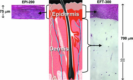
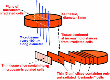
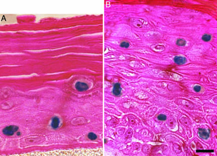
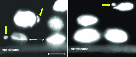
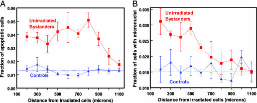
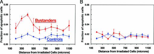
A central tenet in understanding the biological effects of ionizing radiation has been that the initially affected cells were directly damaged by the radiation. By contrast, evidence has emerged concerning "bystander" responses involving damage to nearby cells that were not themselves directly traversed by the radiation. These long-range effects are of interest both mechanistically and for assessing risks from low-dose exposures, where only a small proportion of cells are directly hit. Bystander effects have been observed largely by using single-cell in vitro systems that do not have realistic multicellular morphology; no studies have as yet been reported in three-dimensional, normal human tissue. Given that the bystander phenomenon must involve cell-to-cell interactions, the relevance of such single-cell in vitro studies is questionable, and thus the significance of bystander responses for human health has remained unclear. Here, we describe bystander responses in a three-dimensional, normal human-tissue system. Endpoints were induction of micronucleated and apoptotic cells. A charged-particle microbeam was used, allowing irradiation of cells in defined locations in the tissue yet guaranteeing that no cells located more than a few micrometers away receive any radiation exposure. Unirradiated cells up to 1 mm distant from irradiated cells showed a significant enhancement in effect over background, with an average increase in effect of 1.7-fold for micronuclei and 2.8-fold for apoptosis. The surprisingly long range of bystander signals in human tissue suggests that bystander responses may be important in extrapolating radiation risk estimates from epidemiologically accessible doses down to very low doses where nonhit bystander cells will predominate.


Figures

Fig. 1.
Fig. 1.
The two reconstructed normal human skin tissue systems used here. Shown are the keratinocyte-containing epidermis (EPI-200, Left) and full-thickness skin (EFT-300, Right), consisting of a dermal layer containing fibroblasts and an epidermal layer similar to that in EPI-200, containing keratinocytes.
Fig. 2.
Fig. 2.
Schematic of the irradiation procedure. Each tissue sample consists of an 8-mm-diameter cylinder that is 75 μm (epidermal model, EPI-200) or 700 μm (full-skin model, EFT-300) in height that is microbeam-irradiated along a diameter by α-particles (10 particles every 100 μm along the diameter). The microbeam is <5 μm across, less than one cell diameter, so the plane of irradiated cells is no more than two cells wide. After irradiation, the tissue is fixed and sectioned into 5-μm slices parallel to, and progressively farther from, the irradiated plane of cells.
Fig. 3.
Fig. 3.
Examples of apoptosis in unirradiated bystander cells in artificial human skin systems. Shown are EPI-200 (A) and EFT-300 (B) stained with a DermaTACS apoptosis kit; positive apoptotic cells appear blue. Each slice of tissue shown was >200 μm from the plane of irradiated cells and thus received no direct or scattered radiation exposure. Formalin-fixed, paraffin-embedded, 5-μm-thick histological sections are shown. (Scale bar: 10 μm.)
Fig. 4.
Fig. 4.
Micronuclei observed in unirradiated bystander cells in three-dimensional epidermal tissue (EPI-200) stained with DAPI. Each slice of tissue shown was >200 μm from the irradiated cells and thus received no direct or scattered radiation exposure. The support membrane can be seen at the bottom of each image. The large arrows indicate micronuclei associated with individual cell nuclei. The small arrows show the location of a broken nucleoplasmic bridge, indicating, as expected, a plane of cellular division parallel to the membrane. (Scale bar: 10 μm.)
Fig. 5.
Fig. 5.
Fraction of apoptotic (A) and micronucleated (B) cells in unirradiated bystander cells at different distances from the plane of irradiated cells in a three-dimensional human epidermal skin model (EPI-200). Controls refer to sham irradiations, with conditions otherwise identical. Dotted lines show mean value of control points. Each data point (and SEM) is derived from experiments with three independent tissues.
Fig. 6.
Fig. 6.
Fraction of apoptotic cells in unirradiated keratinocyte layers at different distances from a plane of irradiated cells in a three-dimensional, full-thickness human skin model (EFT-300). Controls refer to sham irradiations, with conditions otherwise identical. Each data point (and SEM) is derived from studies with three independent tissues. (A) Microbeam irradiation of a plane of cells only in the epidermal layer, showing a significant bystander response. (B) Microbeam irradiation of a plane of cells only in the dermal layer, showing no evidence of a bystander response in the unirradiated epidermal layer.

Comment in

  • Effects of ionizing radiation in nonirradiated cells.
    Morgan WF, Sowa MB. Morgan WF, et al. Proc Natl Acad Sci U S A. 2005 Oct 4;102(40):14127-8. doi: 10.1073/pnas.0507119102. Epub 2005 Sep 26. Proc Natl Acad Sci U S A. 2005. PMID: 16186485 Free PMC article. No abstract available.

References

    1. Nagasawa, H. & Little, J. B. (1992) Cancer Res. 52, 6394-6396. - PubMed
    1. Morgan, W. F. (2003) Radiat. Res. 159, 567-580. - PubMed
    1. Zhou, H., Randers-Pehrson, G., Waldren, C. A., Vannais, D., Hall, E. J. & Hei, T. K. (2000) Proc. Natl. Acad. Sci. USA 97, 2099-2104. - PMC - PubMed
    1. Lorimore, S. A. & Wright, E. G. (2003) Int. J. Radiat. Biol. 79, 15-25. - PubMed
    1. Ostreicher, J., Prise, K. M., Michael, B. D., Vogt, J., Butz, T. & Tanner, J. M. (2003) Strahlenther. Onkol. 179, 69-77. - PubMed

Publication types

LinkOut - more resources