. 2005 Sep 14;25(37):8349-58.
doi: 10.1523/JNEUROSCI.1570-05.2005.

Bim is a direct target of a neuronal E2F-dependent apoptotic pathway

Affiliations

Bim is a direct target of a neuronal E2F-dependent apoptotic pathway

Subhas C Biswas et al. J Neurosci. .

Abstract

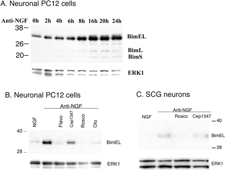
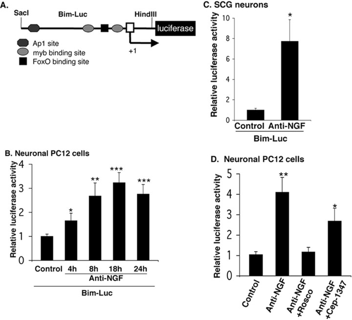
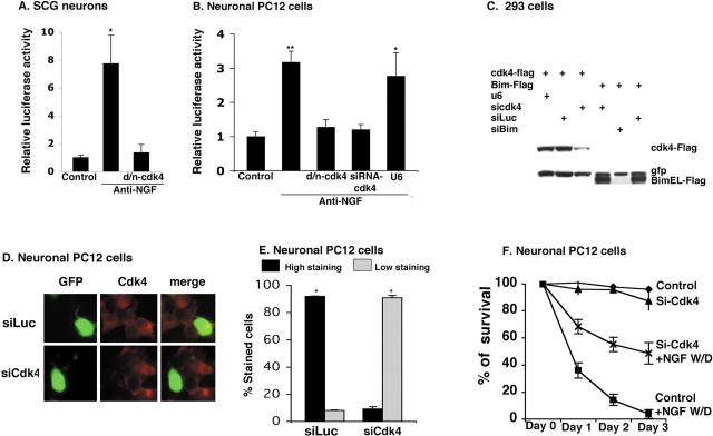
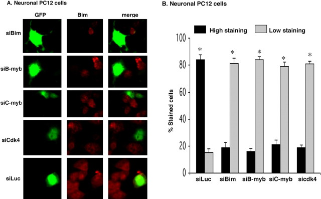
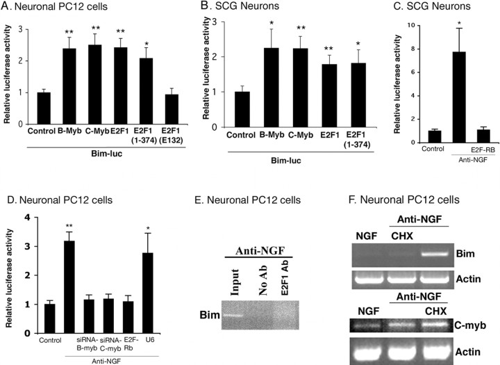
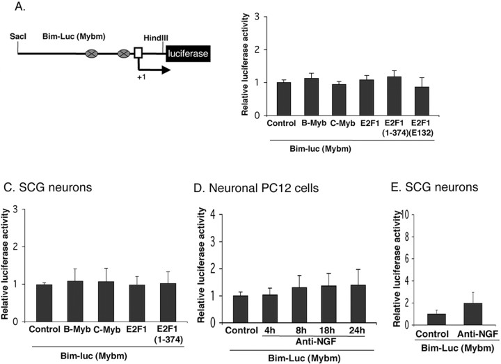
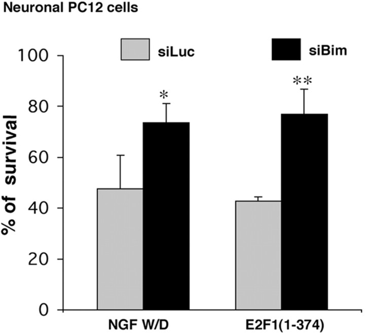
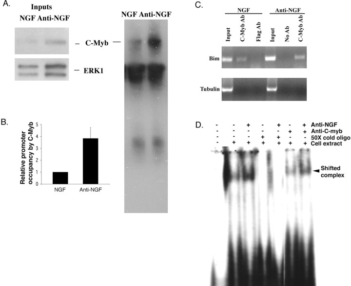
The inappropriate expression/activation of cell-cycle-related molecules is associated with neuron death in many experimental paradigms and human neuropathologic conditions. However, the means whereby this links to the core apoptotic machinery in neurons have been unclear. Here, we show that the pro-apoptotic Bcl-2 homology 3 domain-only molecule Bcl-2 interacting mediator of cell death (Bim) is a target of a cell-cycle-related apoptotic pathway in neuronal cells. Induction of Bim in NGF-deprived cells requires expression and activity of cyclin-dependent kinase 4 (cdk4) and consequent de-repression of E2 promoter binding factor (E2F)-regulated genes including members of the myb transcription factor family. The Bim promoter contains two myb binding sites, mutation of which abolishes induction of a Bim promoter-driven reporter by NGF deprivation or E2F-dependent gene de-repression. NGF deprivation significantly increases endogenous levels of C-myb and its occupancy of the endogenous Bim promoter. These findings support a model in which apoptotic stimuli lead to cdk4 activation, consequent de-repression of E2F-regulated mybs, and induction of pro-apoptotic Bim.


Figures

Figure 1.
Figure 1.
Induction of Bim by NGF withdrawal is completely blocked by cdk inhibitors. A, Time course of the effect of NGF withdrawal on Bim induction. PC12 cells were treated with NGF for 7 d, deprived of NGF for the indicated times, and cell proteins were subjected to Western immunoblotting using enhanced chemiluminescence for the expression of Bim and ERK1. BimL, Bim long; BimS, Bim short. B, The induction of the Bim protein after NGF deprivation is completely blocked by cdk inhibitors and incompletely blocked by inhibition of JNK signaling. PC12 cells were treated with NGF for 7 d and then deprived of NGF for 24 h in presence or absence of 1 μm flavopiridol (Flavo), 200 nm CEP-1347, 20 μm roscovitine (Rosco), or 200 μm olomoucine (Olo). Western immunoblotting was as in A. C, The cdk inhibitor roscovitine blocks Bim upregulation in cultured SCG neurons. Sympathetic neurons, after 3 d of culture, were deprived of NGF for 24 h in presence or absence of 20 μm roscovitine (Rosco) or 200 nm CEP-1347. Western immunoblotting was as in A.
Figure 2.
Figure 2.
The Bim promoter is activated by NGF withdrawal in neuronal PC12 cells and sympathetic neurons. A, Schematic representation of rat Bim promoter-driven luciferase reporter constructs. The Bim–lucreporter consists of ∼3 kb of the Bim rat gene extending 5′ from exon 1 in the pGL3-Basic vector. An arrow indicates the transcription start site (+1). B, Bim promoter-driven luciferase activity is induced after NGF withdrawal in neuronal PC12 cells. Neuronal PC12 cells were transfected with 0.5 μg of Bim–luc reporter and 0.1 μg of the Renilla luciferase expression construct pRL–CMV (to control for transfection efficiency) and were maintained with or without NGF for the indicated times, after which luciferase assays were performed. The data are reported as relative firefly luciferase activity normalized to Renilla luciferase activity and represent means ± SEM of four experiments. Asterisks denote statistically significant differences from control: *p < 0.05; **p < 0.005; ***p < 0.001. C, Bim promoter-driven luciferase activity is increased after NGF withdrawal from sympathetic neurons. Sympathetic neurons were transfected and assayed for luciferase activity as described in B, except that they were maintained with or without NGF for 18 h. The data are means ± SEM of five experiments. The asterisk denotes statistically significant differences from control: *p < 0.004. D, Induction of Bim promoter-driven luciferase activity after NGF withdrawal is completely blocked by a cdk inhibitor and incompletely blocked by inhibition of the JNK pathway. Neuronal PC12 cells were transfected and assayed for luciferase activity as described in B, except that they were treated with 20 μm roscovitine (Rosco) or 200 nm CEP-1347 during 18 h of NGF deprivation. The data are means ± SEM of three experiments. Asterisks denote statistically significant differences from control: *p < 0.04; **p < 0.008.
Figure 3.
Figure 3.
Cdk4 is required for Bim expression. A, B, Induction of Bim promoter-driven luciferase activity after NGF deprivation in cultures of sympathetic neurons (A) and neuronal PC12 cells (B) is blocked by inhibition/loss of cdk4 activity. Cultures were cotransfected with 0.5 μg of Bim–luc and 0.1 μgofthe Renilla luciferase expression construct pRL–CMV with 0.5 μg of empty pCMV vector (Control) or indicated construct, maintained for 24 h, and then deprived of NGF for 18 h, after which luciferase assays were performed. Data were normalized as in Figure 2 and are means ± SEM of three or four experiments. Asterisks denote statistically significant differences from control: A,*p < 0.004; B,*p < 0.001, ** p < 0.0003. C, Cdk4 and Bim siRNAs suppress cdk4 and Bim expression, respectively. HEK 293 cells were cotransfected with pCMS–EGFP expressing Flag–cdk4 or Flag–BimEL and pU6–cdk4–siRNA, pSIREN–Luc–siRNA, pSIREN–Bim–siRNA, or empty pU6 vector. After 48 h, cells were lysed and subjected to Western immunoblotting as described in Figure 1 A with anti-Flag and anti-EGFP as probes. D, E, Cdk4 siRNA reduces endogenous cdk4 expression in neuronal PC12 cells after NGF withdrawal. Neuronal PC12 cells were cotransfected with pSIREN–cdk4–siRNA (siCdk4) or pSIREN–Luc–siRNA (siLuc) and pCMS–EGFP, maintained for 48 h, and then deprived of NGF for 18 h, after which immunostaining with antibodies against cdk4 (red) and GFP (green) was performed. Percentage of stained cells pertains to the proportions of transfected cells (as indicated by staining for GFP) that show cdk4 staining either high (more than that of NGF-maintained cells) or low (equal or less than that of NGF-maintained cells). Data represent means ± SEM of three experiments. Approximately 50 cells were evaluated perculture. Asterisks denote statistically significant differences between low-staining cells and high-staining cells: *p < 0.0001. F, Cdk4 siRNA protects neuronal PC12 cells from death evoked by NGF deprivation. Cells were cotransfected with pCMS–EGFP and either empty pU6 (Control) or pU6–cdk4–siRNA and 24 h later subjected to NGF withdrawal (W/D). Numbers of transfected (green) cells were determined at indicated times, and percentage survival was calculated relative to the number of transfected cells present before exposure to apoptotic conditions. Data represent means ± SEM of three experiments performed in triplicate.
Figure 4.
Figure 4.
siRNAs targeted to Bim, B-myb, C-myb, and cdk4 repress upregulation of endogenous Bim in neuronal PC12 cells subjected to NGF withdrawal. A, B, Neuronal PC12 cells were cotransfected with the indicated constructs and pCMS–EGFP, maintained for 48 h, and then deprived of NGF for 18 h, after which they were immunostained with antibodies against Bim (red) and GFP (green). Percentage of stained cells pertains to the proportions of transfected cells (as indicated by staining for GFP) that show Bim staining either high (more than that of NGF-maintained cells) or low (equal or less than that of NGF-maintained cells). Data represent means ± SEM of three experiments. Approximately 50 cells were evaluated per culture. Asterisks denote statistically significant differences between low-staining cells and high-staining cells: *p < 0.0001.
Figure 5.
Figure 5.
E2F-dependent de-repression of myb is required for induction of Bim promoter activity. Neuronal PC12 cells (A) and sympathetic neurons (B) were cotransfected with 0.5 μg of Bim–luc reporter and 0.1 μg of the Renilla luciferase expression construct pRL–CMV with 0.5 μg of empty pCMV vector (Control), B-myb, C-myb, E2F1, E2F1 (1–374), or E2F1 (E132) and maintained for 24 h, after which luciferase assays were performed. Data were normalized as in Figure 2 and represent means ± SEM for three or four experiments. Asterisks denote statistically significant differences from control: A, *p < 0.006, **p < 0.0001; B, *p < 0.0001, **p < 0.00004. C, D, Induction of Bim promoter-driven luciferase activity after NGF deprivation in cultures of sympathetic neurons (C) and neuronal PC12 cells (D) is blocked by interference with B- and C-myb-promoted gene regulation (D) and repression of E2F responsive genes (C, D). Cultures were cotransfected with 0.5 μg of Bim–luc and 0.1 μg of the Renilla luciferase expression construct pRL–CMV with 0.5μg of empty pCMV vector (Control) or indicated construct, maintained for 24 h, and then deprived of NGF for 18 h, after which luciferase assays were performed. Data were normalized as in Figure 2 and are means ± SEM of three or four experiments. Asterisks denote statistically significant differences from Control: C,*p < 0.004; D, *p < 0.001, **p < 0.0003. E, E2F-responsive elements of the Bim promoter are not occupied in response to NGF deprivation in PC12 cells. PC12 cells were treated with NGF for 6 d and then maintained for an additional day without NGF. Cells were then subjected to ChIP as described in Materials and Methods using anti-E2F1 (Santa Cruz Biotechnology). The immunoprecipitated material was subjected to PCR using Bim primers that flanked one E2F1 binding site. Agarose gel electrophoresis was performed on products of PCR. Templates were DNA derived from cells before ChIP (input), and DNA in immunoprecipitates were derived by ChIP with anti-E2F1. Comparable results were achieved in three independent experiments. F, The protein synthesis inhibitor cycloheximide (CHX) blocks the induction of Bim mRNA expression but not C-myb mRNA expression after NGF deprivation. PC12 cells were treated with NGF for 7 d and then deprived of NGF for 4 h in presence or absence of 10 μg/ml CHX, and total RNA was isolated, reverse transcribed, and amplified by PCR using rat Bim-, rat C-myb-, and actin-specific primers.
Figure 6.
Figure 6.
The activation of Bim promoter by NGF withdrawal or B-myb, C-myb, E2F1, and an E2F truncation mutant lacking a transactivation domain in neuronal PC12 cells and sympathetic neurons requires myb binding sites. A, Schematic representation of rat mutant Bim promoter-driven luciferase reporter constructs. The mutant lacks two myb binding sites shown as ovals. B, C, The activation of Bim promoter by B-myb, C-myb, E2F1, and E2F1(1–374) requires myb binding sites. Neuronal PC12 cells (B) and sympathetic neurons (C) were cotransfected with 0.5 μg of mutant Bim–luc reporter and 0.1 μg of the Renilla luciferase expression construct pRL–CMV with 0.5 μg of empty pCMV vector (Control), B-myb, C-myb, E2F1, E2F1(1–374), or E2F1(E132) and maintained for 24 h, after which luciferase assays were performed. Data were normalized as in Figure 2 and represent means ± SEM for three or four experiments. D, Induction of Bim promoter-driven luciferase activity after NGF withdrawal in neuronal PC12 cells requires myb binding sites. Neuronal PC12 cells were transfected with 0.5 μg of mutant Bim–luc reporter and 0.1 μg of the Renilla luciferase expression construct pRL–CMV and were maintained with or without NGF for the indicated times, after which luciferase assays were performed. The data are reported as relative firefly luciferase activity normalized to Renilla luciferase activity and represent means ± SEM of four experiments. E, Induction of Bim promoter-driven luciferase activity after NGF withdrawal from sympathetic neurons requires myb binding sites. Sympathetic neurons were transfected and assayed for luciferase activity as described in D, except that they were maintained with or without NGF for 18 h. The data are means ± SEM of three experiments.
Figure 7.
Figure 7.
Bim siRNA protects neuronal PC12 cells from death evoked by NGF withdrawal (W/D) and E2F derepression. Neuronal PC12 cells were cotransfected with pCMS–EGFP and pSIREN–Luc–siRNA (siLuc) or pSIREN–Bim–siRNA (siBim) and E2F1 (1–374) as indicated, maintained for 48 h, and then deprived of NGF for 18 h as indicated, after which time numbers of surviving transfected (EGFP+) cells were counted. Data represent means ± SEM of three experiments performed in triplicates. Asterisks denote statistically significant differences from Control (siLuc): *p < 0.05; **p < 0.01.
Figure 8.
Figure 8.
NGF deprivation induces occupation of the endogenous Bim promoter by endogenous C-myb. A, NGF deprivation increases the amount of endogenous C-myb recovered in ChIP assays. PC12 cells were treated with NGF for 6 d and then maintained for an additional day with or without NGF. Equal numbers of cells from each condition were subjected to ChIP as described in Materials and Methods using anti-C-myb. The immunoprecipitated protein–DNA complexes were boiled in sample buffer and analyzed by Western immunoblotting using C-myb antiserum. Comparable results were achieved in an independent experiment. B, NGF withdrawal enhances occupancy of the Bim promoter by C-myb. Neuronal PC12 cells treated as in A were subjected to ChIP with anti-C-myb. The immunoprecipitated material was subjected to real-time quantitative PCR using Bim primers that flanked one C-myb binding site. Data were normalized to the amount of Bim DNA present in the sample before immunoprecipitation (input) as determined by real-time quantitative PCR using the same primers. Comparable results were achieved in four independent experiments. Error bars represent SEM. C, Verification that C-myb occupies the endogenous Bim promoter. Agarose gel electrophoresis was performed on products of quantitative real-time PCR using material derived from neuronal PC12 cells treated as in A as template. Templates were DNA derived from cells before ChIP (input) and DNA in immunoprecipitates derived by ChIP with anti-C-myb or anti-Flag. Primers corresponded to a portion of the Bim promoter as in B (top) or to a portion of theα-tubulin gene (bottom). D, Endogenous C-myb complexes with the Bim promoter in neuronal PC12 cells, and such complexes are increased in response to NGF deprivation. PC12 cells were treated with NGF for 7 d and then maintained for 8 h with or without NGF. Equal amounts of cell extracts from each condition were subjected to electrophoretic mobility gel-shift assays using a rat Bim promoter sequence(lanes 2–7). A50-fold excess of unlabeled probe was included where indicated. Samples in lanes 6 and 7 were incubated with anti-C-myb antibody (Santa Cruz Biotechnology). The solid arrowhead marks the Bim–C-myb complex that is selectively supershifted by anti-C-myb.

References

    1. Angelastro JM, Ignatova TN, Kukekov VG, Steindler DA, Stengren GB, Mendelsohn C, Greene LA (2003) Regulated expression of ATF5 is required for the progression of neural progenitor cells to neurons. J Neurosci 23: 4590–4600. - PMC - PubMed
    1. Bain J, McLauchlan H, Elliott M, Cohen P (2003) The specificities of protein kinase inhibitors: an update. Biochem J 371: 199–204. - PMC - PubMed
    1. Batistatou A, Greene LA (1991) Aurintricarboxylic acid rescues PC12 cells and sympathetic neurons from cell death caused by nerve growth factor deprivation: correlation with suppression of endonuclease activity. J Cell Biol 115: 461–471. - PMC - PubMed
    1. Biswas SC, Greene LA (2002) Nerve growth factor (NGF) down-regulates the Bcl-2 homology 3 (BH3) domain-only protein Bim and suppresses its proapoptotic activity by phosphorylation. J Biol Chem 277: 49511–49516. - PubMed
    1. Bouillet P, Zhang LC, Huang DC, Webb GC, Bottema CD, Shore P, Eyre HJ, Sutherland GR, Adams JM (2001) Gene structure alternative splicing, and chromosomal localization of pro-apoptotic Bcl-2 relative Bim. Mamm Genome 12: 163–168. - PubMed

Publication types

LinkOut - more resources