. 2005 Sep 14;25(37):8375-85.
doi: 10.1523/JNEUROSCI.2164-05.2005.

Neuroprotective mechanisms of lithium in murine human immunodeficiency virus-1 encephalitis

Affiliations

Neuroprotective mechanisms of lithium in murine human immunodeficiency virus-1 encephalitis

Huanyu Dou et al. J Neurosci. .

Abstract

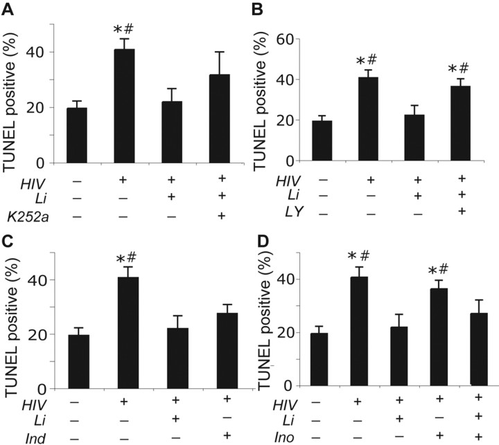
Lithium (Li) has garnered considerable interest as a neuroprotective drug for a broad range of nervous system disorders. Its neuroprotective activities occur as a consequence of glycogen synthase kinase-3beta (GSK-3beta) inhibition leading to downstream blockade of beta-catenin and Tau phosphorylation. In the present study, we investigated Li-mediated neuroprotective mechanisms in laboratory and murine human immunodeficiency virus-1 (HIV-1) encephalitis (HIVE) models. In laboratory tests, Li protected neurons from neurotoxic secretions of HIV-1-infected monocyte-derived macrophages (MDMs). This neuroprotection was mediated, in part, through the phosphatidyl inositol 3-kinase/Akt and GSK-3beta pathways. To examine the effects of Li treatment in vivo, MDMs were injected into the basal ganglia of severe combined immunodeficient mice and then Li was administered (60 mg/kg/d). Seven days after MDM injection, mice were killed and CNS tissue was collected and subjected to immunocytochemical and Western blot assays for leukocyte and neural antigens, GSK-3beta, and key kinase substrates such as beta-catenin and Tau. Numbers of HIV-1 p24 antigen-positive MDMs were unaltered by Li treatment of HIVE mice. Similarly, the greatly increased extent of astrocyte and microglia activation in HIVE mice (10-fold and 16-fold, respectively, compared with unmanipulated controls) was also unaltered by Li. In contrast, Li restored HIVE-associated loss of microtubule-associated protein-2-positive neurites and synaptic density while reducing levels or activity of phospho-Tau Ser202, phospho-beta-catenin, and GSK-3beta. Electrophysiological recordings showed diminished long-term potentiation in hippocampal slices of HIVE mice that were restored by Li. Based on these data, the use of Li as an adjuvant for HIV-1-associated dementia is now being pursued.


Figures

Figure 1.
Figure 1.
Li protects human fetal neurons against toxicity of culture fluids from HIV-1-infected MDMs. Human fetal neurons were cultivated for 2 weeks and then exposed to culture fluids of uninfected (control) and HIV-1-infected MDM [25% (v/v)] for 5 d in the presence or absence of Li (10 μm; as indicated), after which the cultures were analyzed by immunostaining. LY (50 μm), Ind (10 μm), and Ino (10 μm) were added at the time of neuronal exposure to the HIV-1+ fluids. A, Fluorescent images show immunoreactivity for MAP-2 (left, green; right, red), SYP (left, red), and GFAP (right, green). This analysis revealed a high density of dendritic nodes and long neuritic processes with prominent cell bodies and synaptic staining in control neurons. Diminished dendritic nodes, short neurites, and loss of processes were apparent in neurons treated with HIV-1+ fluids and were reversed by Li (right). In addition, extensive astrogliosis (reflected by prominent GFAP immunoreactivity) was detected in all cultures exposed to HIV-1+fluids. Quantitative analysis of these immunostaining results are presented for synaptophysin (B), MAP-2(C), and GFAP (D). SYP expression (p < 0.01) and MAP-2+ neurite length (p < 0.003) were reduced in neurons treated with HIV-1+ fluids compared with untreated control cultures. In Li-treated cultures, the expression of SYP and MAP-2 was essentially restored (p < 0.05 and p < 0.004, respectively) when compared with untreated HIV-1+ neurons. LY added to HIV-1+ fluid-exposed cultures in the presence of Li blocked the neuroprotective effects of Lion SYP and MAP-2 expression (p < 0.05 and p < 0.01, respectively). As a GSK-3 inhibitor, Ind also induced neuroprotective activities, parallel to what was observed when supplied to cells exposed to HIV-1+ fluids (SYP, p < 0.03; MAP-2, p = 0.055). Additional 10 μm Ino added to Li-treated HIV-1+-exposed neurons had no effect on Li neuroprotection. These results are representative of three individual experiments performed in quadruplicate determinations. Original magnification, 400×. The asterisk denotes a statistically significant difference when compared with cells that receive HIV-1+ fluids; the number sign denotes a statistically significant difference compared with cultures that were exposed to HIV-1+ fluids in the presence of Li.
Figure 2.
Figure 2.
Quantitative measures of neuronal apoptosis link specific cell-signaling pathways and Li-mediated neuroprotection. Human fetal neurons were cultivated for 2 weeks and then exposed to culture fluids of HIV-1-infected MDM [25% (v/v)] for 5 d. In the presence or absence of Li (10 mm), LY (50μm), Ind (10μm), K252a (100 nm), and Ino (10μm) were added at the time of neuronal exposure to the HIV-1+ fluids. Uninfected MDM culture fluids served as controls. The levels of neuronal apoptosis were measured by TUNEL with normalization by DAPI staining. Increased apoptosis was observed in neurons treated with HIV-1+ fluids compared with untreated control cultures (p < 0.001). This was reversed by Li (p < 0.003). A, The TrkB inhibitor K252a failed to statistically alter TUNEL staining in all treatment groups. The PI3-kinase/Akt pathway was reflected by LY. B, LY added to Li-treated neurons exposed to HIV-1+ fluids prevented Li-induced reductions in TUNEL staining (p < 0.02). C, The GSK-3β inhibitor Ind had effects similar to those observed for Li. Ind reduced the levels of apoptosis in neurons exposed to HIV-1+ culture fluids when compared with neurons treated with HIV-1 fluids alone (p < 0.05). D, Ino added to Li-treated neurons exposed to HIV-1+ fluids showed similar TUNEL levels as Li administration alone (p = 0.114). These results are representative of three individual experiments performed in quadruplicate determinations. Original magnification, 400×. The asterisk denotes a statistically significant difference when compared in a pairwise manner with untreated control cells that did not receive HIV-1+ fluids; the number sign denotes a statistically significant difference compared with cultures that were exposed to HIV-1+ fluids in the presence of Li.
Figure 3.
Figure 3.
Neuropathological analysis of Li-treated HIVE mice. Serial 5 μm brain sections, cut through the needle track, were immunostained for vimentin, HIV-1 p24, GFAP, and lectin. In all cases, the chromagen used to reveal the immunostained cells is brown. Thirty fields in 15 sections were collected from the injection site through the BG and cerebral cortex and subjected to quantitative morphometric analysis. No differences were detected in numbers of human cells (vimentin staining), HIV-1 p24 antigen, astrocyte reactivity (GFAP staining), or microglial activation (lectin staining) in the HIVE mice versus the HIVE mice that were exposed to Li (as shown by the representative microscope fields; immunostaining data not shown). Original magnification, 200×.
Figure 4.
Figure 4.
Li protects neurons in HIVE mice. Immunostaining was performed on formalin-fixed paraffin-embedded brain tissue sections from the BG of HIVE mice using antibodies specific to MAP-2 (green) and vimentin (red). Spatial relationships between neurites (MAP-2+) and human MDM (vimentin positive) were determined by superimposing the image data (Merge). In HIVE animals, loss of MAP-2 immunostaining (green) was most intense around the implanted human MDM (red) but also extended beyond this region. In contrast, Li-treated HIVE mice showed significantly stronger MAP-2 immunostaining throughout the tissue, even in regions in which HIV-1-infected MDMs were present (as revealed by the orange staining in the merged image). Original magnification, 200×.
Figure 5.
Figure 5.
Li protects neuronal dendrites and synaptic clefts. A, Immunostaining was performed on serial 5μm formalin-fixed paraffin-embedded brain tissue sections with antibodies to MAP-2 (a marker for dendrites; purple stain, top panels) and SYP (a marker for synaptic clefts; brown stain, bottom panels). B, Quantitation of SYP immunostaining also revealed a significant loss of SYP+ synapses in the BG HIVE mice (filled bars), relative to untreated control mice (open bars) (p < 0.03). This loss of SYP+ staining density in the HIVE mice was restored by treatment with Li (gray bars). C, Quantitative analysis of the immunostained sections revealed that there was a significant loss of MAP-2+ dendrites in the BG and cortex of HIVE mice (filled bars) relative to untreated control mice (open bars) (p < 0.002 for BG; p < 0.003 for cortex, respectively). Li treatment partially restored MAP-2 levels in both sites in the HIVE mice (gray bars; p < 0.02 and p < 0.05 in the BG and in the cortex). D, E, To confirm histological results for MAP-2 levels, brain tissue protein was analyzed by quantitative Western blot assays. Data were normalized by β-tubulin. HIVE mice showed lower levels of MAP-2 compared with control (p < 0.05). Li treated to HIVE animals showed an increase in MAP-2 levels. Original magnification, 200×. The asterisk denotes a statistically significant difference when compared with sham mice; the number sign denotes a statistically significant difference compared with Li-treated HIVE mice.
Figure 6.
Figure 6.
HIVE inflammatory responses diminish expression of a cell-surface marker associated with newly generated neurons within the hippocampus; this is reversed by Li. A, Brain tissue from control or HIVE mice (with or without Li treatment) was prepared and subjected to immunostaining using antibodies specific for MAP-2 or PSA-NCAM, which is a marker for newly generated neurons within the hippocampus. The representative images reveal significant MAP-2+ neuronal loss within the CA1 region and DG of hippocampi in HIVE mice, as well as loss of PSA-NCAM+ cells. This was confirmed by quantitative analysis of immunostained sections. B, The length of MAP-2+ dendrites (Index) was significantly decreased in the CA1 (p < 0.003) and DG (p < 0.03) hippocampi subregions of HIVE mice compared with sham-operated mice. MAP-2+ neurite length was restored to normal in the Li-treated HIVE mice (HIV-Li). A significant increase of MAP-2+ neurites was shown in the CA1 (p < 0.05) and DG (p<0.03) of HIV-Li animals compared with HIVE mice. C, The numbers of PSA-NCAM+ cells in the DG of HIVE mice were reduced, relative to sham controls (p < 0.0002). Li restored PSA-NCAM+ cell numbers to normal levels. D, E, Not only were the total number of PSA-NCAM+ cells reduced in HIVE mice, average numbers of dendrites on these cells were also reduced relative to sham controls (p < 0.001), as was the average length of dendrites (p < 0.01). Treatment with Li increased both PSA-NCAM+ dendrite numbers (p < 0.01) and length (p < 0.03) in the HIVE mice. Original magnification, 400×. The asterisk denotes a statistically significant difference when compared with sham mice; the number sign denotes a statistically significant difference when compared with Li-treated HIVE mice.
Figure 7.
Figure 7.
Diminished LTP in both the ipsilateral and contralateral hippocampi after injection of HIV-1-infected MDM into the left BG. A illustrates the magnitude of LTP recorded from hippocampal brain slices from sham-operated animals (control), HIVE SCID mice (HIV), and HIV mice treated with Li (HIV+Li). B shows LTP measured 70 min after HFS. No significant difference (p > 0.05) was observed between the two hemispheres. Diminished LTP levels were recorded in HIVE mice when compared with sham mice (p < 0.05); Li treatment significantly improved this functional measure in these animals (p < 0.05). The asterisk denotes a statistically significant difference when compared with sham mice; the number sign denotes a statistically significant difference when compared with Li-treated HIVE mice.
Figure 8.
Figure 8.
Pathways for Li-mediated neuroprotection in HIVE mice. A, Western blot assays were performed on tissue extracts using antibodies specific for GSK-3β, phosphorylated β-catenin, β-catenin, Tau (all isoforms; Tau5 antibody), and two phosphorylated isoforms of Tau (Ser202 and Thr181). B, C, Data were quantitated densitometrically. As expected, expression levels of GSK-3β were unaltered in all groups of mice. Total levels of β-catenin were slightly reduced in the HIVE mice relative to sham controls, whereas levels of phosphorylated β-catenin were significantly upregulated in the HIVE mice (p < 0.05). This is consistent with a rise in the enzymatic activity of GSK-3β in the HIVE animals. Li treatment essentially reversed these effects on β-catenin. C, Quantitative analysis of the Tau phosphorylation at residues 202 and 181 was performed by measuring immunoreactivity with phospho-specific antisera directed against the Tau202 and Tau181 residues and then normalizing the resulting densitometric data in terms of total Tau immunoreactivity (determined using the Tau5 antibody, which recognizes all forms the protein). This analysis revealed increases in both Ser202 (p < 0.05) and Thr181 (p < 0.01) phosphorylated isoforms of Tau in HIVE mice (HIV), compared with control animals (sham). These changes were reversed by Li treatment of HIVE mice. The asterisk denotes a statistically significant difference when compared with sham mice; the number sign denotes a statistically significant difference when compared with Li-treated HIVE mice.
Figure 9.
Figure 9.
Engagement of GSK-3β and β-catenin phosphorylation in Li-mediated neuroprotection. A, Culture fluids from HIV-1-infected MDM [25% (v/v)] were added to human fetal neurons in the presence or absence of 10 mm Li and the indicated compounds (LY and Ind) for 2 h. Neuronal cell lysates were then prepared and subjected to Western blot assays using antibodies specific for total GSK-3β, phospho-serine9 GSK-3β, β-catenin, and phospho-serine33,37-β-catenin. B, C, Li reduced Ser9 phosphorylation of GSK-3β. Quantitative analysis of immunoblots performed from neuronal lysates demonstrated that total GSK-3β levels were not statistically different in untreated cells versus cells exposed to HIV-1+ fluids, regardless of the presence of Li. B, In contrast, phosphorylation of GSK-3β Ser9 was decreased after 5 d of neuronal exposure to HIV-1+ culture fluids when compared with replicate neurons treated with culture fluids from uninfected MDMs (control; p < 0.01) and Li-treated cells exposed to HIV-infected materials (p < 0.03). C, When LY, in combination with Li, was exposed to neurons treated with HIV-1+ fluids, GSK-3β Ser9 phosphorylation was reduced compared with replicate neuronal cultures given Li (p < 0.05) or with negative controls (p < 0.02). GSK-3β phosphorylation was also increased by Ind (p < 0.05). D, E, Li reducedβ-catenin phosphorylation. Totalβ-catenin levels (D) were unaltered by exposure to HIV-1+ fluids or treatment with Li. HIV-1+ culture fluids did, however, induce significant levels of phospho-serine33,37-β-catenin, relative to control culture fluids (p < 0.03 and p < 0.02). These levels were reduced by Li (p < 0.05) and negated when LY was added to cells with Li (p < 0.05 and p < 0.05), compared with cells treated with control fluids or HIV-1+ fluids in the presence of Li. Finally, Ind diminished the phosphorylation of β-catenin at Ser33,37 regardless of the addition of Li. These results are representative of three individual experiments done in quadruplicate determinations (the asterisk denotes a statistically significant difference when compared in a pairwise manner to untreated control cells that did not receive HIV-1+ fluids; the number sign denotes a statistically significant difference compared with cultures that were exposed to HIV-1+ fluids in the presence of Li).

References

    1. Anderson ER, Boyle J, Zink WE, Persidsky Y, Gendelman HE, Xiong H (2003) Hippocampal synaptic dysfunction in a murine model of human immunodeficiency virus type 1 encephalitis. Neuroscience 118: 359–369. - PubMed
    1. Anderson ER, Gendelman HE, Xiong H (2004) Memantine protects hippocampal neuronal function in murine human immunodeficiency virus type 1 encephalitis. J Neurosci 24: 7194–7198. - PMC - PubMed
    1. Berry GT, Buccafusca R, Greer JJ, Eccleston E (2004) Phosphoinositide deficiency due to inositol depletion is not a mechanism of lithium action in brain. Mol Genet Metab 82: 87–92. - PubMed
    1. Bhat RV, Budd Haeberlein SL, Avila J (2004) Glycogen synthase kinase 3: a drug target for CNS therapies. J Neurochem 89: 1313–1317. - PubMed
    1. Bodner A, Toth PT, Miller RJ (2004) Activation of c-Jun N-terminal kinase mediates gp120IIIB- and nucleoside analogue-induced sensory neuron toxicity. Exp Neurol 188: 246–253. - PubMed

Publication types

MeSH terms