. 2005 Sep 14;25(37):8567-77.
doi: 10.1523/JNEUROSCI.2493-05.2005.

Loss of Mtmr2 phosphatase in Schwann cells but not in motor neurons causes Charcot-Marie-Tooth type 4B1 neuropathy with myelin outfoldings

Affiliations

Loss of Mtmr2 phosphatase in Schwann cells but not in motor neurons causes Charcot-Marie-Tooth type 4B1 neuropathy with myelin outfoldings

Annalisa Bolis et al. J Neurosci. .

Abstract

Mutations in MTMR2, the myotubularin-related 2 gene, cause autosomal recessive Charcot-Marie-Tooth type 4B1 (CMT4B1). This disorder is characterized by childhood onset of weakness and sensory loss, severely decreased nerve conduction velocity, demyelination in the nerve with myelin outfoldings, and severe functional impairment of affected patients, mainly resulting from loss of myelinated fibers in the nerve. We recently generated Mtmr2-null(neo) mice, which show a dysmyelinating neuropathy with myelin outfoldings, thus reproducing human CMT4B1. Mtmr2 is detected in both Schwann cells and neurons, in which it interacts with discs large 1/synapse-associated protein 97 and neurofilament light chain, respectively. Here, we specifically ablated Mtmr2 in either Schwann cells or motor neurons. Disruption of Mtmr2 in Schwann cells produced a dysmyelinating phenotype very similar to that of the Mtmr2-null(neo) mouse. Disruption of Mtmr2 in motor neurons does not provoke myelin outfoldings nor axonal defects. We propose that loss of Mtmr2 in Schwann cells, but not in motor neurons, is both sufficient and necessary to cause CMT4B1 neuropathy. Thus, therapeutical approaches might be designed in the future to specifically deliver the Mtmr2 phospholipid phosphatase to Schwann cells in affected nerves.


Figures

Figure 1.
Figure 1.
Generation of mice with Mtmr2 conditional ablation. A, First row, Genomic structure of Mtmr2 encompassing exon 4 (the wild-type Mtmr2 locus). Second row, Targeting construct in which Mtmr2 genomic fragments are represented by thick lines and vector sequences are represented by thin lines (the targeting vector). Third row, Mtmr2 locus after homologous recombination in ES cells as reported by Bolino et al. (2004) (the Mtmr2Flneo allele). Fourth row, Targeted allele without the neo cassette, removed in vivo using the Flpe transgene (the Mtmr2Fl allele). Fifth row, Targeted allele without neo and exon 4 (the Mtmr2-null allele). FRT, Flp recognition target; H, HindIII; K, KpnI; TK, thymidine kinase gene. B, PCR on genomic DNA from different tissues of Mtmr2Fl/nullneo//P0-Cre mice performed using primers A+E. Exon 4 was nearly completely excised in the sciatic nerve. C, PCR on genomic DNA from different tissues of Mtmr2Fl/nullneo//HB9-Cre mice performed using primers A+E. Exon 4 was nearly completely excised in the pancreas (as shown in previous studies) and only partially in the spinal cord (sp. cord), in which other neuronal cell types, endothelial and glial cells, are located. No recombination was noted in other tissues. D, PCR on genomic DNA from laser capture-microdissected motor neurons (MN) of Mtmr2-nullneo mice. The primer pair A+D was used. In these mice, exon 4 was removed from both Mtmr2-targeted alleles in which the neo cassette is still present. D', PCR on genomic DNA from laser capture-microdissected motor neurons (MN) of Mtmr2Fl/nullneo//HB9-Cre mice. The primer pair A+E was used. In these mice, exon 4 was absent in motor neurons from the spinal cord (MN), and a band of ∼300 bp was detected. If exon 4 was present, a band of ∼800 bp (containing exon 4 and one loxP site) would have been amplified, as in the case of the non-recombined tissues in B and C. Empty lanes (–) in D and D' correspond to the negative control of the PCRs. E, In situ mRNA hybridization analysis using, as a probe, a portion of the Mtmr2 cDNA-containing exons 11–14. Mtmr2 mRNA is detected in motor neurons from wild-type (wt) and Mtmr2Fl/nullneo//P0-Cre mice but not in Mtmr2Fl/nullneo//HB9-Cre or Mtmr2-nullneo motor neurons.
Figure 2.
Figure 2.
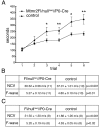
Behavioral and electrophysiological analysis of Mtmr2Fl/nullneo//P0-Cre mice. A, Rotarod test analysis performed on mutant and control animals at 6 months of age. For statistical analysis, the StatView program (version 5.0) with the paired t test method was used. *p < 0.05; **p < 0.01. B, Electrophysiological analysis of Mtmr2Fl/nullneo//P0-Cre mice and controls at 6 months of age. Reduction of NCV and prolonged F-wave latencies in mutant mice are comparable with those obtained for Mtmr2-nullneo mice at the same age, as reported by Bolino et al. (2004). C, Electrophysiological analysis of Mtmr2Fl/nullneo//P0-Cre mice and controls at 12 months of age.
Figure 3.
Figure 3.
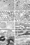
Morphological analysis of Mtmr2Fl/nullneo//P0-Cre mice. A–D, Semithin analysis of sciatic nerves from mutant mice in a transverse section at 7 weeks and at 6, 12, and 15 months. Arrowheads indicate myelin outfoldings and redundant loops. Arrows indicate remyelinated axons at older ages. E, F, Digital nerves (plantar) from normal (E) and mutant (F) mice at 12 months of age. G, H, Semithin analysis of sciatic nerves in a longitudinal section at 12 months of age. Myelin infoldings (H) and outfoldings (G), which arise at Schmidt–Lanterman incisures, are indicated by an asterisk. H, Myelin outfoldings also arise at paranodal regions. NR, Node of Ranvier. I, J, Electron microscopy of a sciatic nerve analyzed at 12 months of age showing myelin outfoldings at paranodal regions (I), which appeared normal with axo-glial junctions and adherens junctions in register (arrow) (J, enlarged). K, L, Electron microscopy showing myelin outfoldings at Schmidt–Lanterman incisures (arrows), enlarged in L. In the outfold, beneath the compact myelin sheath, Schwann cell cytoplasm containing vesicles and axoplasm contiguous with the main myelinated axon might be observed. Scale bar: A–F, 25 μm; G, H, 6 μm; I, 2 μm; J, 400 nm; K, 3.5 μm; L, 1.5 μm.
Figure 4.
Figure 4.
Morphological analysis of Mtmr2-nullneo mice at 12 and 15 months of age. A–E, Different shape of myelin outfoldings in Mtmr2-nullneo mouse sciatic nerves at older ages when the complexity of the dysmyelinating phenotype progresses. In A and D, upto five satellite loops are noted around a main myelinated fiber, which also appear around the nucleus (D). C, Myelin outfoldings in small caliber-myelinated fibers are shown. E, A thin myelinated fiber is depicted. F, G, Semithin analysis in a longitudinal section of mutant sciatic nerves at 12 months of age. Asterisks indicate myelin loops arising at Schmidt–Lanterman incisures. H–J, Teased fiber analysis of Mtmr2-nullneo sciatic nerves at 6 months of age. Myelin outfoldings originate at paranodal regions but also at Schmidt–Lanterman incisures (asterisk). NR, Node of Ranvier. Scale bar: (in J) A–E, 30 μm; F–J, 8 μm.
Figure 5.
Figure 5.
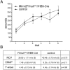
Behavioral and electrophysiological analysis of Mtmr2Fl/nullneo//HB9-Cre mice. A, Rotarod test analysis performed on mutant and control animals at 6 months of age. No significant differences were observed in motor performances using the accelerating rod method. B, Electrophysiological analysis of Mtmr2Fl/nullneo//HB9-Cre mice and controls at 6 months of age. NCV, F-wave latencies, and CMAP registered in mutant animals were similar to controls.
Figure 6.
Figure 6.
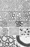
Morphological analysis of Mtmr2Fl/nullneo//HB9-Cre mice. A–D, Semithin analysis of sciatic nerves from mutants at 1 month (A), 2 months (B), 6 months (C), and 12 months (D) of age. No myelin outfoldings were noted. E–G, Quadriceps nerves from mutant mice at 5 months (E), 8 months (F), and 12 months (G) of age and a saphenous nerve at 5 months of age (H) in which no myelin outfoldings were observed. I, Digital nerve at 12 months without signs of dysmyelination (plantar nerve with ∼40 myelinated fibers). J, K, ATPase isozyme staining of posterior compartment muscles of the leg shows that no type grouping occurred. L, M, Electron microscopy of quadriceps nerves at 12 months of age in which compact-myelin structure appears normal, and no axonal swelling was noted in the axoplasm. Scale bar: (in M) A–I, 25 μm; J, K, 80 μm; L, 8 μm; M, 3.5 μm.
Figure 7.
Figure 7.
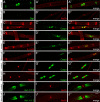
Expression analysis of nodal, paranodal, and juxtaparanodal markers in Mtmr2-nullneo sciatic nerves. Immunohistochemistry was performed on teased fibers from Mtmr2-nullneo and normal sciatic nerves at 2 months of age. The following stainings were performed: Dlg1, on normal (A–A“) and mutant (B–B”) fibers. Double labeling of Dlg1 (green) and NaCh channel clusters (red), which mark the node of Ranvier, was performed. Dlg1 is almost absent from paranodal loops and flanking incisures in Mtmr2-nullneo fibers (B–B“). MAG, on normal (C–C”) and mutant (D–D“) fibers. Double labeling of MAG (red) and Caspr (green), which mark paranodal regions on the axolemma, was performed. MAG staining is reduced in paranodes from null fibers (compare the merge in C” and D“). Reduced stainings of both Dlg1 and MAG in Mtmr2-nullneo fibers were further confirmed using quantification of the average signal intensity. E-cadherin (E-cadh) on normal (E–E”) and mutant (F–F“) fibers, double labeled with E-cadh (red) and Caspr (green), is shown. No difference in signal intensity and distribution was detected between normal and mutant fibers (compare the merge in E” with F“). NaCh clusters on normal (G–G”) and mutant (H–H“) fibers are shown. Double labeling of NaCh (red) and Caspr (green) was performed. Kv1.1/1.2 clusters (green) on normal (I–I”) and mutant (J–J“) fibers costained with NaCh (red) are shown. No difference in staining intensity or distribution of NaCh, Caspr, and Kv1.1/1.2 clusters was appreciated between normal and mutant fibers. Scale bar: (in J”) A–F, 10 μm; G–J”, 5 μm.
Figure 8.
Figure 8.
A, Analysis of protein clusters at nodal, paranodal, and juxtaparanodal regions of Mtmr2-nullneo sciatic nerves. B, Na cluster size (wild type, 1.25 ± 0.04μm; knock-out, 1.46 ± 0.04 μm average) was normalized using the Na cluster diameter for each node (wild type, 1.85 ± 0.05 μm; knock-out, 2.04 ± 0.06 μm average); n, Number of nodes analyzed. Caspr cluster length (wild type, 3.80 ± 0.09μm; knock-out, 4.06 ± 0.09μm average) was normalized using Caspr diameter (wild type, 2.59 ± 0.09μm; knock-out, 2.57 ± 0.07μm average). n refers to paranodes analyzed. Kv1.1/1.2 cluster length (wild type, 8.22 ± 0.47 μm; knock-out, 6.91 ± 0.40 μm average) was normalized with the Na cluster diameter (wild type, 2.75 ± 0.09 μm; knock-out, 2.48 ± 0.11 μm average). n refers to juxtaparanodes analyzed. Kv1.1/1.2 cluster distance (wild type, 6.47 ± 0.33 μm; knock-out, 6.54 ± 0.29 μm average) was normalized using Na cluster diameter as for Kv1.1/1.2 cluster length. n is the number of fibers analyzed. The difference in Kv1.1/1.2 cluster distance between mutant and normal fibers was not statistically significant.
Figure 9.
Figure 9.
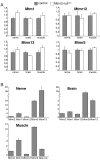
Expression analysis of MTMR family members. A, The expression of Mtm1, Mtmr12, Mtmr13, and Mtmr5 mRNAs was determined by RT-PCR in nerve, brain, and muscle from Mtmr2-nullneo and wild-type animals. TaqMan probes (Applied Biosystems) and the comparative CT method (ABI PRISM Sequence Detection System User Bulletin 2; Applied Biosystems) were used. mRNA expression levels (2–ΔΔCT values) in mutant tissues are indicated as n-folds with respect to the levels in normal tissues, taken as 1. Diagrams show the average of expression levels calculated on at least three animals per genotype in muscle and brain and, for the nerve, on at least three separate pools of two animals each. SD was calculated for triplicate samples of each reaction, and SEM is indicated on the media of the determinations from different animals. B, mRNA expression levels of Mtmr2, Mtm1, Mtmr12, Mtmr5, and Mtmr13 in normal nerve, brain, and muscle. Mtm1, Mtmr12, Mtmr5, and Mtmr13 levels are indicated relative to Mtmr2, taken as 1. The mRNA expression levels of different genes were compared at the same CT value, using the same endogenous references.

References

    1. Antonellis A, Ellsworth RE, Sambuughin N, Puls I, Abel A, Lee-Lin SQ, Jordanova A, Kremensky I, Christodoulou K, Middleton LT, Sivakumar K, Ionasescu V, Funalot B, Vance JM, Goldfarb LG, Fischbeck KH, Green ED (2003) Glycyl tRNA synthetase mutations in Charcot-Marie-Tooth disease type 2D and distal spinal muscular atrophy type V. Am J Hum Genet 72: 1293–1299. - PMC - PubMed
    1. Arber S, Han B, Mendelsohn M, Smith M, Jessell TM, Sockanathan S (1999) Requirement for the homeobox gene HB9 in the consolidation of motor neuron identity. Neuron 23: 659–674. - PubMed
    1. Azzedine H, Bolino A, Taieb T, Birouk N, Di Duca M, Bouhouche A, Benamou S, Mrabet A, Hammadouche T, Chkili T, Gouider R, Ravazzolo R, Brice A, Laporte J, LeGuern E (2003) Mutations in MTMR13, a new pseudophosphatase homologue of MTMR2 and Sbf1, in two families with an autosomal recessive demyelinating form of Charcot-Marie-Tooth disease associated with early-onset glaucoma. Am J Hum Genet 72: 1141–1153. - PMC - PubMed
    1. Bolino A, Muglia M, Conforti FL, LeGuern E, Salih MA, Georgiou DM, Christodoulou K, Hausmanowa-Petrusewicz I, Mandich P, Schenone A, Gambardella A, Bono F, Quattrone A, Devoto M, Monaco AP (2000) Charcot-Marie-Tooth type 4B is caused by mutations in the gene encoding myotubularin-related protein-2. Nat Genet 25: 17–19. - PubMed
    1. Bolino A, Marigo V, Ferrera F, Loader J, Romio L, Leoni A, Di Duca M, Cinti R, Cecchi C, Feltri ML, Wrabetz L, Ravazzolo R, Monaco AP (2002) Molecular characterization and expression analysis of Mtmr2, mouse homologue of MTMR2, the Myotubularin-related 2 gene, mutated in CMT4B. Gene 283: 17–26. - PubMed

Publication types

MeSH terms

Substances