. 2005 Sep 15;437(7057):417-21.
doi: 10.1038/nature03928.

WNT7b mediates macrophage-induced programmed cell death in patterning of the vasculature

Affiliations

WNT7b mediates macrophage-induced programmed cell death in patterning of the vasculature

Ivan B Lobov et al. Nature. .

Abstract

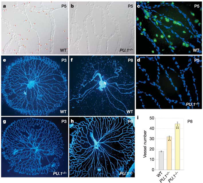
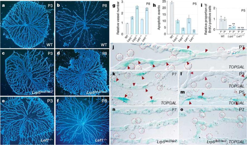
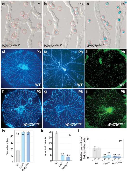
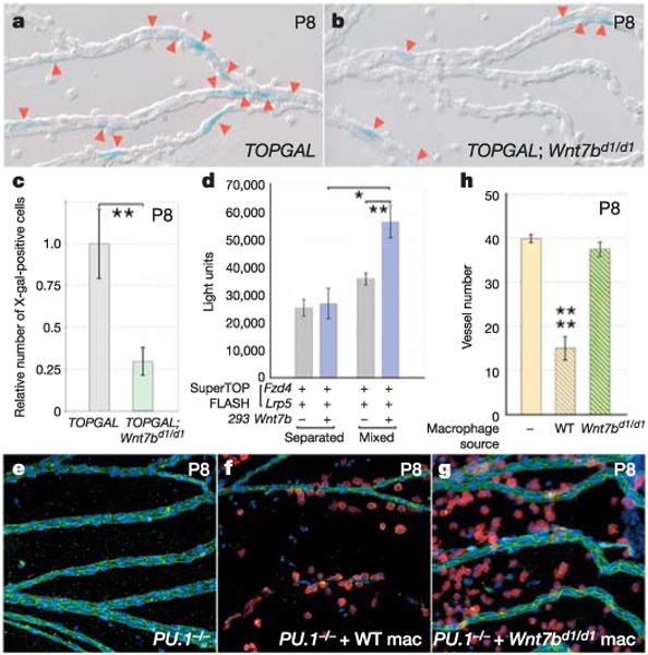
Macrophages have a critical role in inflammatory and immune responses through their ability to recognize and engulf apoptotic cells. Here we show that macrophages initiate a cell-death programme in target cells by activating the canonical WNT pathway. We show in mice that macrophage WNT7b is a short-range paracrine signal required for WNT-pathway responses and programmed cell death in the vascular endothelial cells of the temporary hyaloid vessels of the developing eye. These findings indicate that macrophages can use WNT ligands to influence cell-fate decisions--including cell death--in adjacent cells, and raise the possibility that they do so in many different cellular contexts.


Conflict of interest statement

The authors declare no competing financial interests.

Figures

Figure 1
Figure 1. Regression of the hyaloid vessels is macrophage-dependent
a–d, Hyaloid vessel preparations from wild-type (a, c) and PU.1−/− (b, d) mice at P5. a, b, Differential interference contrast illumination indicating the presence of macrophages in wild-type mice (a, with red dots adjacent) and the absence of macrophages in PU.1−/− mice (b). c, d, Fluorescent immunostaining for macrophages (F4/80, a macrophage-specific marker, green) and nuclei (blue). e–h, Hyaloid vessels from wild-type (e, f) and PU.1−/− (g, h) animals of the indicated ages stained with Hoechst 33258. i, Hyaloid vessel number in wild-type, PU.1−/− and PU.1−/− mice at P8. All error bars are standard errors. Significance levels: three asterisks, 0.0001 < P < 0.001; four asterisks, P < 0.0001. Original magnification: ×50 (e–h); ×400 (a–d). WT, wild type.
Figure 2
Figure 2. The WNT pathway response in VECs is required for hyaloid vessel regression
a–f, Hoechst-33258-labelled hyaloid vessel preparations from mice of the indicated ages and genotypes. g–i, Vessel number at P8 (g), number apoptotic events at P5 (h) and BrdU labelling index in primary (1°) and secondary (2°) capillary branches at P5 (i) in the hyaloid vessels of mice with the indicated genotypes. Lrp5−/− is an abbreviated reference to the Lrp5lacZ/lacZ allele. j–o, X-gal staining (blue) in hyaloid vessels from mice of the indicated ages and genotypes. Intensely stained TOPGAL-expressing cells (red arrowheads) and resident macrophages (red dashed circles) are apparent. All error bars are standard errors. Significance levels: two asterisks, 0.001 < P < 0.01; three asterisks, 0.0001 < P < 0.001; four asterisks, P < 0.0001. Original magnification: ×50 (a–f); ×630 (k–o); ×1,000 (j). WT, wild type.
Figure 3
Figure 3. WNT7b is required for hyaloid vessel regression and is expressed in macrophages
a–c, X-gal staining (blue) of hyaloid vessels from Wnt7b+/lacZ mice of the indicated ages. Macrophages are indicated by dashed red circles. d–g,i,j, Hyaloid vessels from wild-type (d,e,i) and Wnt7bd1/d1 (f,g,j) animals at the indicated ages, stained with Hoechst 33258 (d–g) or anti-F4/80 (i, j). h, k, l, Quantification, in hyaloid vessels from mice with the indicated genotypes, of vessel number at P8 (h), apoptotic events at P5 (k) and BrdU labelling index in primary (1°) and secondary (2°) capillary branches at P5 (l). All error bars are standard errors. Significance levels: two asterisks, 0.001 < P < 0.01. Original magnification: ×50 (d–g, i, j); ×630 (a–c). WT, wild type.
Figure 4
Figure 4. Macrophages are a critical paracrine source of WNT7b required for hyaloid vessel regression
a, b, X-gal staining (blue) of P8 hyaloid vessel preparations in TOPGAL (a) and TOPGAL; Wnt7bd1/d1 (b) mice. Red arrowheads indicate stained cells. c, Relative number of X-gal-stained cells in TOPGAL (grey bar) and TOPGAL;Wnt7bd1/d1 (green bar) hyaloid preparations at P8. The data are normalized for the increased vessel number of the Wnt7bd1/d1 mutants. d, Quantification of WNT responsiveness in SuperTOPFLASH cells transfected with Fzd4 and Lrp5 and either mixed with WNT7b producer cells or separated from them by restricting SuperTOPFLASH responder cells and WNT7b producer cells to each half of a culture dish. e–g, Hyaloid vessel preparations at P8 from uninjected PU.1−/− mice (e), PU.1−/− mice injected with wild-type macrophages (f) and PU.1−/− mice injected with Wnt7bd1/d1 macrophages (g). Preparations are labelled for the F4/80 marker (red), vascular endothelial cell cadherin (green) and nuclei (blue). Original magnification: ×200. h, Vessel number at P8 in uninjected PU.1−/− mice (yellow bar), PU.1−/− mice injected with wild-type macrophages (yellow/grey bar) and PU.1−/− mice injected with Wnt7bd1/d1 macrophages (yellow/green bar). All error bars are standard errors. Significance levels: one asterisk, 0.01 < P < 0.05; two asterisks, 0.001 < P < 0.01; four asterisks, P < 0.0001. WT, wild type; Mac, macrophages.

References

    1. Savill J, Dransfield I, Gregory C, Haslett C. A blast from the past: clearance of apoptotic cells regulates immune responses. Nature Rev Immunol. 2002;2:965–975. - PubMed
    1. Lang RA, Bishop MJ. Macrophages are required for cell death and tissue remodeling in the developing mouse eye. Cell. 1993;74:453–462. - PubMed
    1. Diez-Roux G, Lang RA. Macrophages induce apoptosis in normal cells in vivo. Development. 1997;124:3633–3638. - PubMed
    1. Hoeppner DJ, Hengartner MO, Schnabel R. Engulfment genes cooperate with ced-3 to promote cell death in Caenorhabditis elegans. Nature. 2001;412:202–206. - PubMed
    1. Reddien PW, Cameron S, Horvitz HR. Phagocytosis promotes programmed cell death in C. elegans. Nature. 2001;412:198–202. - PubMed

Publication types