Comparative Study
. 2005 Oct 5;24(19):3369-79.
doi: 10.1038/sj.emboj.7600811. Epub 2005 Sep 15.

Dual-mode recognition of noncanonical tRNAs(Ser) by seryl-tRNA synthetase in mammalian mitochondria

Affiliations
Comparative Study

Dual-mode recognition of noncanonical tRNAs(Ser) by seryl-tRNA synthetase in mammalian mitochondria

Sarin Chimnaronk et al. EMBO J. .

Abstract

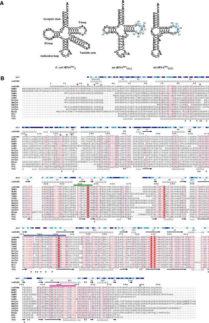
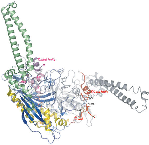
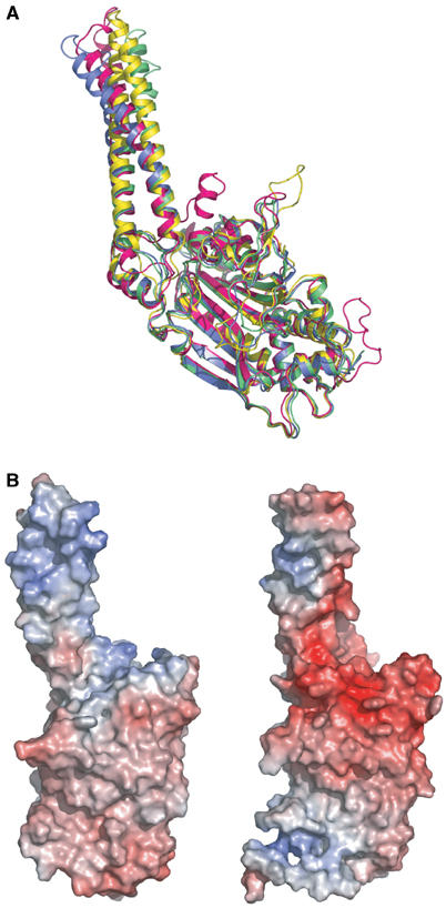
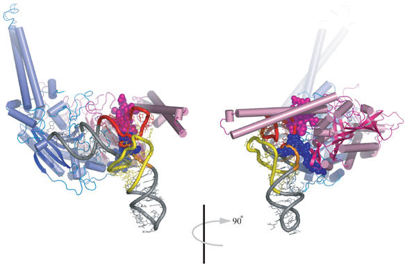
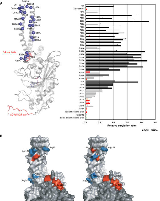
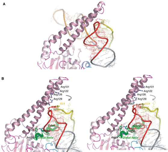
The secondary structures of metazoan mitochondrial (mt) tRNAs(Ser) deviate markedly from the paradigm of the canonical cloverleaf structure; particularly, tRNA(Ser)(GCU) corresponding to the AGY codon (Y=U and C) is highly truncated and intrinsically missing the entire dihydrouridine arm. None of the mt serine isoacceptors possesses the elongated variable arm, which is the universal landmark for recognition by seryl-tRNA synthetase (SerRS). Here, we report the crystal structure of mammalian mt SerRS from Bos taurus in complex with seryl adenylate at an atomic resolution of 1.65 A. Coupling structural information with a tRNA-docking model and the mutagenesis studies, we have unraveled the key elements that establish tRNA binding specificity, differ from all other known bacterial and eukaryotic systems, are the characteristic extensions in both extremities, as well as a few basic residues residing in the amino-terminal helical arm of mt SerRS. Our data further uncover an unprecedented mechanism of a dual-mode recognition employed to discriminate two distinct 'bizarre' mt tRNAs(Ser) by alternative combination of interaction sites.


Figures

Figure 1
Figure 1
Unique structural characteristics of the tRNA/synthetase system for serine in mammalian mitochondria. (A) Secondary-structure diagrams of typical and atypical tRNAsSer are depicted in cloverleaf representations. The left panel shows the canonical serine isoacceptor from E. coli possessing an elongated variable arm as a discriminative tRNA identity for recognition by SerRS. This identity element disappears in both mammalian mt tRNAsSer, as shown in the middle and right panels for B. taurus mt tRNASerUGA and tRNASerGCU, respectively. Identity elements shifting to the T-loop of mt tRNAsSer as previously reported (Shimada et al, 2001) are illustrated in blue characters for the tRNA mutagenesis studies, and indicated by arrowheads for tRNA-footprinting experiments. (B) The amino-acid sequence alignments of SerRS from three domains of life. The organisms referred to in the sequences alignment and corresponding accession numbers of the Swiss Protein Data Base are as follows. The recombinant mt SerRS from B. taurus used for structure determination (reBvMt) having a hexahistidine tag in the N-terminus is in the top row and is numbered. A red arrowhead indicates the N-terminus of the mature protein in mitochondria. The secondary structure elements as defined by the crystal structure are shown above the sequence and labeled according to bacterial SerRS (Cusack et al, 1990). Solvent accessibility (acc) is shown in blue, cyan and white for accessible, intermediate and buried residues, respectively. Another five mt SerRSs from Homo sapiens (HmMt; Q9NP81), Mus musculus (MuMt; Q9JJL8), Drosophila melanogaster (DmMt), Caenorhabditis elegans (CeMt) and Saccharomyces cerevisiae (ScMt; P38705) are shown, followed by five eukaryotic cytoplasmic from B. taurus (BvCyt), H. sapiens (HmCyt; P49591), D. melanogaster (DmCyt), C. elegans (CeCyt; Q18678) and S. cerevisiae (ScCyt; P07284), then follows one archaeal from Methanococcus jannaschii (Mjan; Q58477), and two bacterial from E. coli (Eco; P09156) and T. thermophilus (Tth; P34945). The secondary structure elements of T. thermophilus are also shown below the sequence (Fujinaga et al, 1993). Conserved amino acids are in white in red-filled rectangles. Similar residues are in red, surrounded by blue lines. Class II synthetase motifs 1, 2 and 3 are underlined in green, purple and pink, respectively. Residues involved in tRNA recognition are indicated with asterisks for mt SerRS and with sharps for T. thermophilus SerRS, as suggested by mutational (in this study) and crystallographic studies (Biou et al, 1994), respectively.
Figure 2
Figure 2
Ribbon representation of the three-dimensional crystal structure of B. taurus mt SerRS at 1.65 Å resolution, depicted in different colors for three α-helical bundles and β-sheet strands. The mitochondria-unique N-terminal distal helix (of monomer 2) and C-tail (of monomer 1) are colored in red, with the stick representations showing the mutual interaction sites. The second monomer is drawn as gray ribbons for clarity. Secondary structure elements are denominated corresponding to bacterial SerRS (Cusack et al, 1990). This figure and subsequent figures were composed using PyMol (http://pymol.sourceforge.net/).
Figure 3
Figure 3
Distinct features of B. taurus mt SerRS compared with bacterial SerRS. (A) Superposition of Cα traces of mt SerRS with the known structures of bacterial SerRSs reveals characteristic insertions in both N- and C-terminus. B. taurus mt SerRS is colored in magenta, whereas SerRS from T. thermophilus and E. coli are in blue and yellow, respectively. The second monomer of T. thermophilus SerRS in the tRNASer complex (green) reveals the alteration of the orientation and curvature of the helical arm during tRNA recognition (Biou et al, 1994). (B) Solvent-accessible surfaces of T. thermophilus (left panel) and mt (right panel) SerRS are represented in the same orientation corresponding to Figure 5A, and colored according to electrostatic potential (red for negative and blue for positive), suggesting the interaction sites with the phosphate backbone of tRNA, which differ substantially between the two molecules.
Figure 4
Figure 4
Docking model of yeast tRNAPhe onto mt SerRS. Overall view with the protein shown in ribbon representation is colored in blue and pink for monomers 1 and 2, respectively. The intact tRNA is depicted in gray, except for the D-arm (yellow), variable arm (orange) and the T-arm (red). Seryl adenylates in the active sites are rendered in stick. Mitochondria-specific insertions in the N-terminus of monomer 1 and C-terminus of monomer 2 are depicted in spacefill representations. The right panel is a 90° rotated view clearly revealing steric clashes with the elbow region of tRNA.
Figure 5
Figure 5
Mutagenesis studies of SerRS variants. (A) In all, 12 deletion and 23 alanine-substituted mutants of mt SerRS illustrated in the left panel, and a single mutant of E. coli SerRS with the mt N-terminal distal helix and C-tail inserted, were assayed by in vitro aminoacylation (shown in the right panel) with two native mt serine isoacceptors: tRNAsSerUGA (empty bar) and tRNAsSerGCU (filled bar). The results indicate the initial rate of serylation relative to the WT mt enzyme. Mutations retaining the serylation ability comparable to the WT are colored in black, whereas mutations that lower the serylation rate to <60% of the WT are shown in gray, and mutants that lose the serylation ability (<20%) are in red. (B) Residues involved in recognition of mt tRNASerGCU (left panel) and tRNASerUGA (right panel) are mapped onto the surface of the dimeric structure (light and dark gray for each monomer). The effective sites are colored in cyan, while the critical residues are depicted in red.
Figure 6
Figure 6
Proposed discriminating model of the SerRS–tRNASer complex. (A) illustrates the protein-sandwich model based on the crystal structure of T. thermophilus SerRS–tRNASer (Biou et al, 1994; Cusack et al, 1996). Proteins and tRNAs are colored corresponding to Figure 4. (B) Stereo view of the mt discriminating complex suggests a dissimilar RNA-sandwich model in which three separate parts of the protein are encompassing the T-loop of the modeled tRNA. The mitochondria-unique extensions are shown in green ribbon and tube for the distal helix and the C-tail, respectively. Residues involved in RNA–protein interactions are drawn in stick representations. Gray arrows indicate a likely movement of the helical arm upon tRNA binding to make interactions with the T-loop of tRNA.

References

    1. Achsel T, Gross HJ (1993) Identity determinants of human tRNASer: sequence elements necessary for serylation and maturation of a tRNA with a long extra arm. EMBO J 12: 3333–3338 - PMC - PubMed
    1. Belrhali H, Yaremchuk A, Tukalo M, Larsen K, Berthet-Colominas C, Leberman R, Beijer B, Sproat B, Als-Nielsen J, Grubel G, Legrand JF, Lehmann M, Cusack S (1994) Crystal structures at 2.5 angstrom resolution of seryl-tRNA synthetase complexed with two analogs of seryl adenylate. Science 263: 1432–1436 - PubMed
    1. Bilokapic S, Korencic D, Soll D, Weygand-Durasevic I (2004) The unusual methanogenic seryl-tRNA synthetase recognizes tRNASer species from all three kingdoms of life. Eur J Biochem 271: 694–702 - PubMed
    1. Biou V, Yaremchuk A, Tukalo M, Cusack S (1994) The 2.9 A crystal structure of T. thermophilus seryl-tRNA synthetase complexed with tRNASer. Science 263: 1404–1410 - PubMed
    1. Brunger AT, Adams PD, Clore GM, DeLano WL, Gros P, Grosse-Kunstleve RW, Jiang JS, Kuszewski J, Nilges M, Pannu NS, Read RJ, Rice LM, Simonson T, Warren GL (1998) Crystallography & NMR system: a new software suite for macromolecular structure determination. Acta Crystallogr D 54 (Part 5): 905–921 - PubMed

Publication types

LinkOut - more resources