

The human relaxin receptor (LGR7): expression in the fetal membranes and placenta
- PMID: 16165207
- PMCID: PMC1455164
- DOI: 10.1016/j.placenta.2005.07.011
The human relaxin receptor (LGR7): expression in the fetal membranes and placenta
Abstract
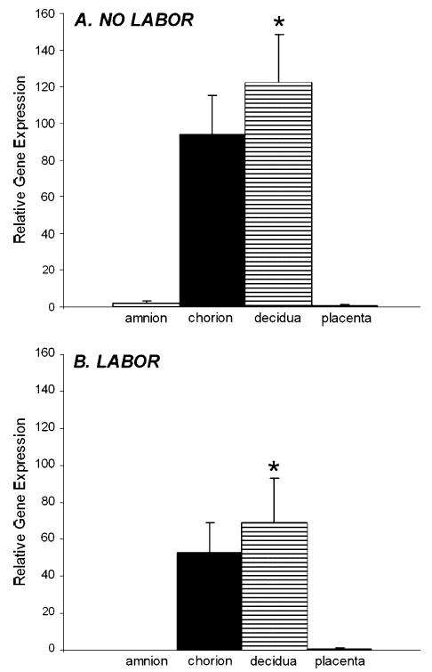
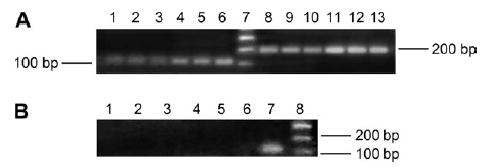
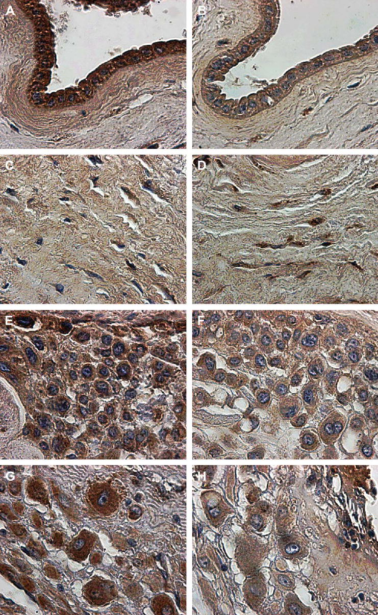
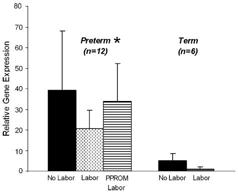
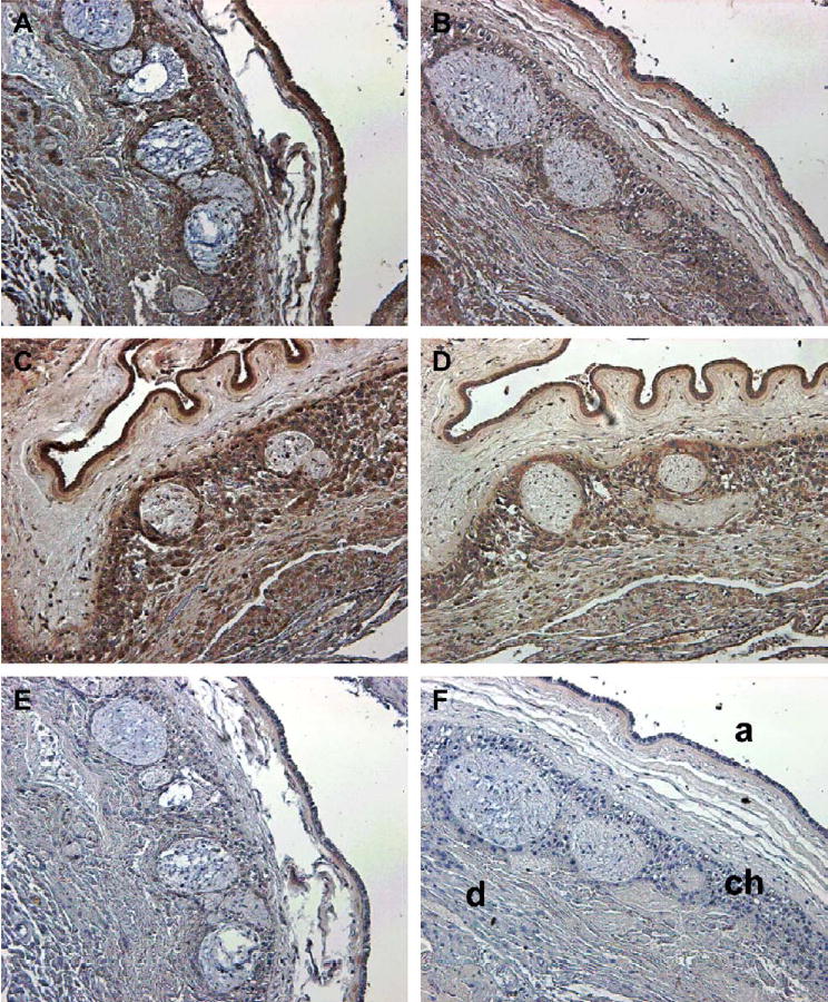
The relaxin receptor has been recently described as a leucine-rich repeat G-protein coupled receptor and designated as LGR7. A closely related receptor, LGR8, is co-expressed by some cells. This study explored the expression of the genes for these receptors in the human fetal membranes and placenta by RT-PCR and the LGR7 protein by immunolocalization. The results showed that LGR7 was well expressed in the fetal membranes, with significantly more in the decidua (p<0.05) than in the amnion. On the other hand, relatively low levels were expressed in the placenta. The major splice variant of LGR7 was undetectable in either the placenta or fetal membranes. Expression of LGR8 was also below the level of detectability in either tissue. Immunostaining for LGR7 was conducted with antisera to both its endodomain and ectodomain, in order to seek evidence for a solubilized ectodomain. However, similar staining patterns were obtained with both antisera, with predominant staining in the cells of the amniotic epithelium, chorionic cytotrophoblast and decidua. Full-thickness fetal membranes from preterm deliveries, before and after labor or after preterm premature rupture of the membrane (PPROM) and labor were collected. In addition, membranes at term, both before and after spontaneous labor were used for analysis of LGR7 gene expression. There was significantly greater LGR7 expressed (p=0.01) in the preterm period compared to term, indicating a potentially important role for relaxin at this time. There was a marginal decline in LGR7 gene expression after labor and delivery both at preterm and term, which did not reach significance. Immunostaining patterns showed less inter-patient variability than did gene expression, with more intense staining for LGR7 after labor and delivery.
Figures
Similar articles
-
Human decidual relaxin and preterm birth.Ann N Y Acad Sci. 2005 May;1041:338-44. doi: 10.1196/annals.1282.054. Ann N Y Acad Sci. 2005. PMID: 15956731
-
Cloning, expression, and functional characterization of relaxin receptor (leucine-rich repeat-containing g protein-coupled receptor 7) splice variants from human fetal membranes.Endocrinology. 2008 Mar;149(3):1277-94. doi: 10.1210/en.2007-1348. Epub 2007 Dec 13. Endocrinology. 2008. PMID: 18079195 Free PMC article.
-
Developmental regulation of the human relaxin genes in the decidua and placenta: overexpression in the preterm premature rupture of the fetal membranes.Biol Reprod. 1997 Oct;57(4):908-20. doi: 10.1095/biolreprod57.4.908. Biol Reprod. 1997. PMID: 9314597
-
Relaxin and the human fetal membranes.Reprod Sci. 2007 Dec;14(8 Suppl):42-5. doi: 10.1177/1933719107310821. Reprod Sci. 2007. PMID: 18089609 Review.
-
Fetal Membranes, Not a Mere Appendage of the Placenta, but a Critical Part of the Fetal-Maternal Interface Controlling Parturition.Obstet Gynecol Clin North Am. 2020 Mar;47(1):147-162. doi: 10.1016/j.ogc.2019.10.004. Epub 2019 Dec 18. Obstet Gynecol Clin North Am. 2020. PMID: 32008665 Review.
Cited by
-
Evidence for existence of insulin-like factor 3 (INSL3) hormone-receptor system in the ovarian corpus luteum and extra-ovarian reproductive organs during pregnancy in goats.Cell Tissue Res. 2021 Jul;385(1):173-189. doi: 10.1007/s00441-021-03410-1. Epub 2021 Feb 15. Cell Tissue Res. 2021. PMID: 33590284
-
Identification of relaxin-responsive cells in the human choriodecidua at term.Ann N Y Acad Sci. 2009 Apr;1160:136-7. doi: 10.1111/j.1749-6632.2008.03786.x. Ann N Y Acad Sci. 2009. PMID: 19416174 Free PMC article.
-
Alternative splicing of G protein-coupled receptors: physiology and pathophysiology.Cell Mol Life Sci. 2009 Oct;66(20):3337-52. doi: 10.1007/s00018-009-0093-4. Epub 2009 Jul 23. Cell Mol Life Sci. 2009. PMID: 19629391 Free PMC article. Review.
-
mRNA and Protein Expression in Human Fetal Membrane Cells: Potential Biomarkers for Preterm Prelabor Rupture of the Fetal Membranes?Int J Mol Sci. 2023 Oct 31;24(21):15826. doi: 10.3390/ijms242115826. Int J Mol Sci. 2023. PMID: 37958809 Free PMC article.
-
Relaxin family peptide receptors Rxfp1 and Rxfp2: mapping of the mRNA and protein distribution in the reproductive tract of the male rat.Reprod Biol Endocrinol. 2007 Jul 10;5:29. doi: 10.1186/1477-7827-5-29. Reprod Biol Endocrinol. 2007. PMID: 17623071 Free PMC article.
References
-
- Hsu SY, Kudo M, Chen T, Nakabayashi K, Bhalla A, van der Spek PJ, et al. The three subfamilies of leucine-rich repeat-containing G protein-coupled receptors (LGR7): identification of LGR6 and LGR7 and the signaling mechanism for LGR7. Mol Endocrinol. 2000;14:1257–71. - PubMed
-
- Hsu SY, Nakabayashi K, Nishi S, Kumagai J, Kudo M, Sherwood OD, et al. Activation of orphan receptors by the hormone relaxin. Science. 2002;295:671–4. - PubMed
-
- Kumagai J, Hsu SY, Mtsumi H, Roh JS, Fu P, Hsueh AJW. INSL3/Leydig insulin-like peptide activates the LGR8 receptor important in testis descent. J Biol Chem. 2002;277:31283–6. - PubMed
-
- Hudson P, Haley M, Cronk M, Crawford R, Haralambidis J, Tregear G, et al. Structure of a genomic clone encoding biologically active human relaxin. Nature. 1983;301:628–31. - PubMed
Publication types
MeSH terms
Substances
Grants and funding
- T34 GM007684-23/GM/NIGMS NIH HHS/United States
- RR1A1-03061/RR/NCRR NIH HHS/United States
- P20 RR011091/RR/NCRR NIH HHS/United States
- P20 RR011091-11/RR/NCRR NIH HHS/United States
- P20 RR011091-060002/RR/NCRR NIH HHS/United States
- T34 GM007684/GM/NIGMS NIH HHS/United States
- R01 HD024314/HD/NICHD NIH HHS/United States
- G12 RR003061-19/RR/NCRR NIH HHS/United States
- RR-11091/RR/NCRR NIH HHS/United States
- U54 RR014607-05/RR/NCRR NIH HHS/United States
- HD-24314/HD/NICHD NIH HHS/United States
- R01 HD024314-12/HD/NICHD NIH HHS/United States
- GM 07684/GM/NIGMS NIH HHS/United States
LinkOut - more resources
Full Text Sources
Molecular Biology Databases
