

Methamphetamine-induced striatal apoptosis in the mouse brain: comparison of a binge to an acute bolus drug administration
- PMID: 16165214
- PMCID: PMC2896282
- DOI: 10.1016/j.neuro.2005.05.014
Methamphetamine-induced striatal apoptosis in the mouse brain: comparison of a binge to an acute bolus drug administration
Abstract
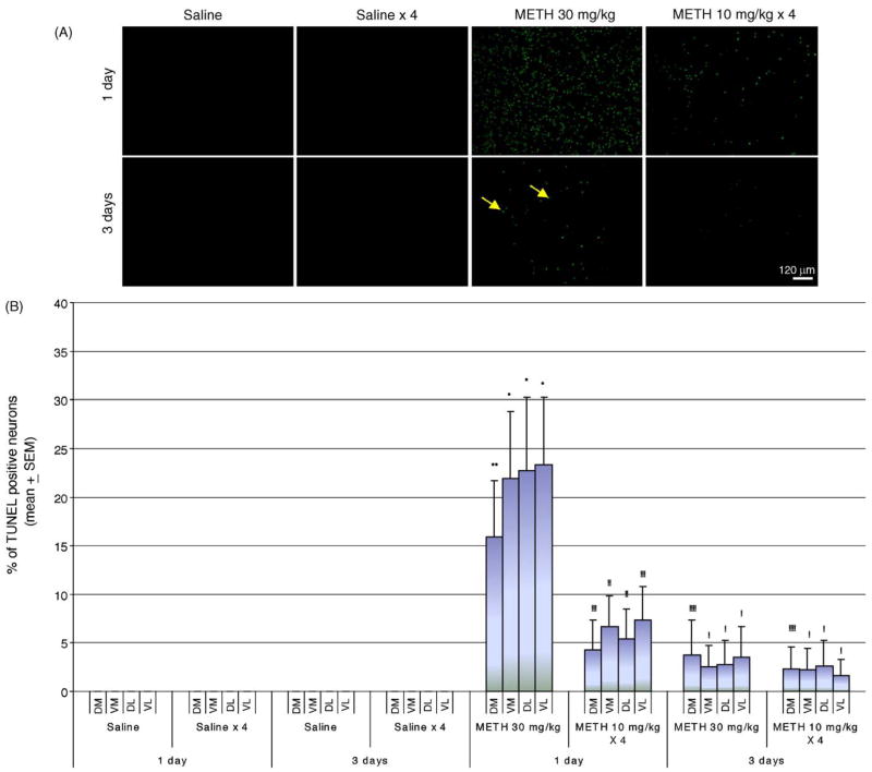
Methamphetamine (METH) is a psychostimulant that induces neural damage in experimental animals and humans. A binge (usually in the 5-10 mg/kg dose range 4 x at 2 h intervals) and the acute bolus drug administration (20-40 mg/kg) of METH have been employed frequently to study neurotoxicity in the brain. In this study we have compared these drug delivery schedules to determine their efficacy to induce striatal apoptosis. Exposure of male mice to a binge of METH at 10mg/kg 4x at 2 h intervals (cumulative dose of 40 mg/kg) was approximately four times less effective in inducing apoptotic cell death (TUNEL staining) 24 h after METH treatment in the striatum than a single bolus administration of 30 mg/kg of METH. The residual TUNEL staining observed three days after METH treatment is proportionately equivalent between a binge and the acute bolus drug administration. Interestingly, a binge of METH induces a hyperthermic response of longer duration. This study demonstrates that an acute bolus drug administration of METH is more effective inducing striatal apoptosis in mice, and therefore, is more suitable for studies assessing the impact of METH on sites post-synaptic to the striatonigral dopamine terminals.
Figures
 ) Saline-1 day, (
 ) Saline × 4–1 day, (
 ) Saline-3 days, (
 ) Saline × 4–3 days, (
 ) METH 30 mg/kg-1 day, (
 ) METH 10 mg/kg × 4–1 day, (
 ) METH 30 mg/kg-3 days, (●) METH 30 mg/kg-3 days. n = 10 for all saline groups. n = 13 for METH treatment groups at day 1. n = 14 for METH treatment groups at day 3.
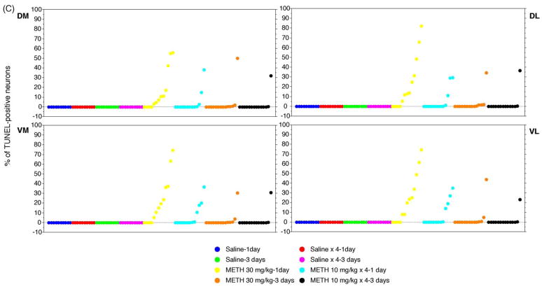 ) Saline-1 day, () Saline × 4–1 day, () Saline-3 days, () Saline × 4–3 days, () METH 30 mg/kg-1 day, () METH 10 mg/kg × 4–1 day, () METH 30 mg/kg-3 days, (●) METH 30 mg/kg-3 days. n = 10 for all saline groups. n = 13 for METH treatment groups at day 1. n = 14 for METH treatment groups at day 3.
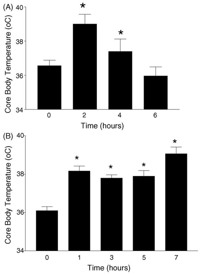
References
-
- Bowyer JF, Harris AJ, Delongchamp RR, Jakab RL, Miller DB, Little AR, et al. Selective changes in gene expression in cortical regions sensitive to amphetamine during the neurodegenerative process. Neurotoxicology. 2004;25:555–72. - PubMed
-
- Davidson C, Gow AJ, Lee TH, Ellinwood EH. Methamphetamine neurotoxicity: necrotic and apoptotic mechanisms and relevance to human abuse and treatment. Brain Res Brain Res Rev. 2001;36:1–22. - PubMed
-
- Deng X, Wang Y, Chou J, Cadet JL. Methamphetamine causes widespread apoptosis in the mouse brain: evidence from using an improved TUNEL histochemical method. Brain Res Mol Brain Res. 2001;93:64–9. - PubMed
-
- Eisch AJ, Marshall JF. Methamphetamine neurotoxicity: dissociation of striatal dopamine terminal damage from parietal cortical cell body injury. Synapse. 1998;30:433–45. - PubMed
-
- Ernst T, Chang L, Leonido-Yee M, Speck O. Evidence for long-term neurotoxicity associated with methamphetamine abuse: a 1H MRS study. Neurology. 2000;54:1344–9. - PubMed
Publication types
MeSH terms
Substances
Grants and funding
LinkOut - more resources
Full Text Sources
Medical
