. 2005 Sep 27;102(39):14086-91.
doi: 10.1073/pnas.0506774102. Epub 2005 Sep 15.

Connections of thalamic modulatory centers to the vocal control system of the zebra finch

Affiliations

Connections of thalamic modulatory centers to the vocal control system of the zebra finch

Eugene Akutagawa et al. Proc Natl Acad Sci U S A. .

Abstract

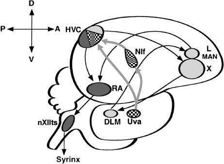
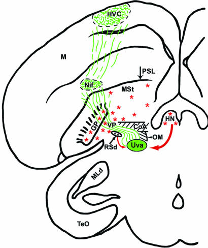
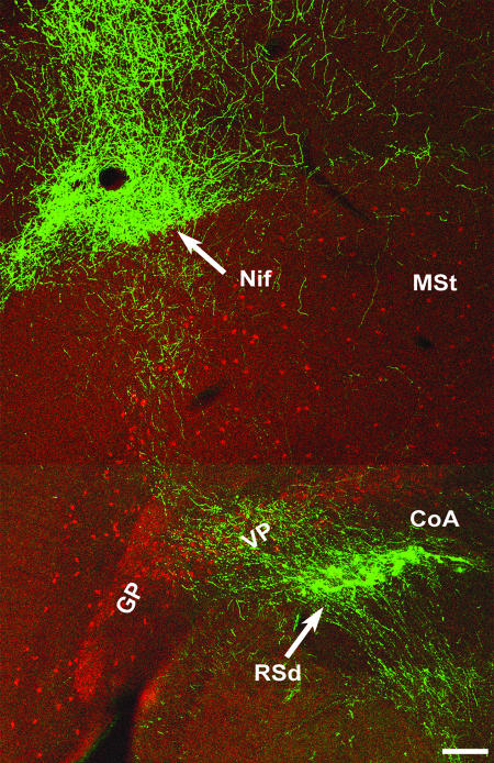
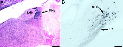
The vocal control system of zebra finches shows auditory gating in which neuronal responses to the individual bird's own song vary with behavioral states such as sleep and wakefulness. However, we know neither the source of gating signals nor the anatomical connections that could link the modulatory centers of the brain with the song system. Two of the song-control nuclei in the forebrain, the HVC (used as the proper name) and the interfacial nucleus of the nidopallium, both show auditory gating, and they receive input from the uvaeform nucleus (Uva) in the thalamus. We used a combination of anterograde and retrograde tracing methods to show that the dorsal part of the reticular formation and the medial habenula (MHb) project to the Uva. We also show by choline acetyl transferase immunohistochemistry that the MHb is cholinergic and sends cholinergic fibers to the Uva. Our findings suggest that the Uva might serve as a hub to coordinate neuromodulatory input into the song system.


Figures

Fig. 1.
Fig. 1.
Schematic representation of the zebra finch brain in sagittal view. The song system can be subdivided into the anterior forebrain circuit (light gray) and the motor pathway (dark gray) and is bridged by their common connection to the HVC, which receives efferent projections from both the Uva and NIf (patterned). nXIIts, tracheosyringeal part of the hypoglossal nucleus; X, area X within the songbird medial striatum; LMAN, lateral magnocellular nucleus of the anterior nidopallium; RA, robust nucleus of the arcopallium; DLM, nucleus dorsolateralis anterior thalami, pars medialis
Fig. 2.
Fig. 2.
A coronal composite rendition summarizing the results of a BDA tracer injection into the Uva. Anterograde-labeled axons (green) from the Uva are shown entering the telencephalon just above the RSd, overlapping with the VP area, and spreading out to innervate both the NIf and HVC. Red arrows show the input areas to the Uva (RSd and HN), and the red asterisks denote the general location of ChAT-IR cells that were relevant to the present study. TeO, optic tectum; MLd, nucleus mesencephalicus lateralis, pars dorsalis; M, mesopallium; HN, habenula nuclei, which include both the lateral habenula and MHb, but only the latter contains ChAT-IR cells; PSL, pallial-subpallial lamina; GP, globus pallidus; OM, occipitomesencephalic tract; MSt, medial striatum; CoA, anterior commisure
Fig. 3.
Fig. 3.
Double fluorescent-labeled anterograde fibers from the Uva (green) and ChAT-IR cells (red) in coronal section. The low-power image shows the general trajectory of the pathway from the Uva, which sends projections to the NIf and keeps coursing upward to ultimately innervate the HVC (compare with Fig. 2). (Scale bar, 200 μm.)
Fig. 4.
Fig. 4.
The MHb in coronal sections. (A) Neutral red and ChAT-IR (dark blue) cells show the location of the MHb between the lateral habenula on the left (lateral) and the anterior-most portion of the cerebellum on the right. Dorsal is up. (Scale bar, 200 μm.) (B) Retrograde-labeled cells from tracer injections into the ipsilateral Uva show labeled axons in the fasciculus retroflexus and cells in the MHb. Dorsal is up, and medial is to the right. (Scale bar, 50 μm.)
Fig. 5.
Fig. 5.
Anterograde transport to the Uva after a BDA injection into the MHb. (A) Low-power coronal section reveals labeled fibers from the MHb innervating the Uva. (Scale bar, 50 μm.) (B) Higher magnification of the same section, which was later counterstained with neutral red. Dorsal is up, and lateral is to the right. (Scale bar, 10 μm.)
Fig. 6.
Fig. 6.
The MHb provides cholinergic input to the Uva. (A) Coronal section of an adult male zebra finch shows ChaT-IR in the Uva. Dorsal is up, and lateral is to the left. (Scale bar, 200 μm.) (B) Low-power view of the MHb double labeled with ChAT-IR (red) and retrograde cells from an ipsilateral Uva injection of a fluorescent green tracer. Dorsal is up, and medial is to the right. (Scale bar, 50 μm.) (C) High magnification of cells double labeled in the MHb. (C1) ChaT-IR cells in the MHb; arrow points to a cell of interest. (C2) The same cell is retrogradely labeled from the Uva with a green tracer. (C3) An overlay of C1 and C2 confirms that this cell is double labeled. Dorsal is up, and medial is to the right. (Scale bar, 20 μm.)

References

    1. Nottebohm, F., Stokes, T. M. & Leonard, C. M. (1976) J. Comp. Neurol. 165, 457-486. - PubMed
    1. Margoliash, D. M. (1983) J. Neurosci. 3, 1039-1057. - PMC - PubMed
    1. Doupe, A. J. & Konishi, M. (1991) Proc. Natl. Acad. Sci. USA 88, 11339-11343. - PMC - PubMed
    1. Coleman, M. J. & Mooney, R. (2004) J. Neurosci. 24, 7251-7256. - PMC - PubMed
    1. Schmidt, M. F. & Konishi, M. (1998) Nat. Neurosci. 1, 513-518. - PubMed

Publication types

Substances

LinkOut - more resources