

Hox genes specify vertebral types in the presomitic mesoderm
- PMID: 16166377
- PMCID: PMC1221883
- DOI: 10.1101/gad.338705
Hox genes specify vertebral types in the presomitic mesoderm
Abstract
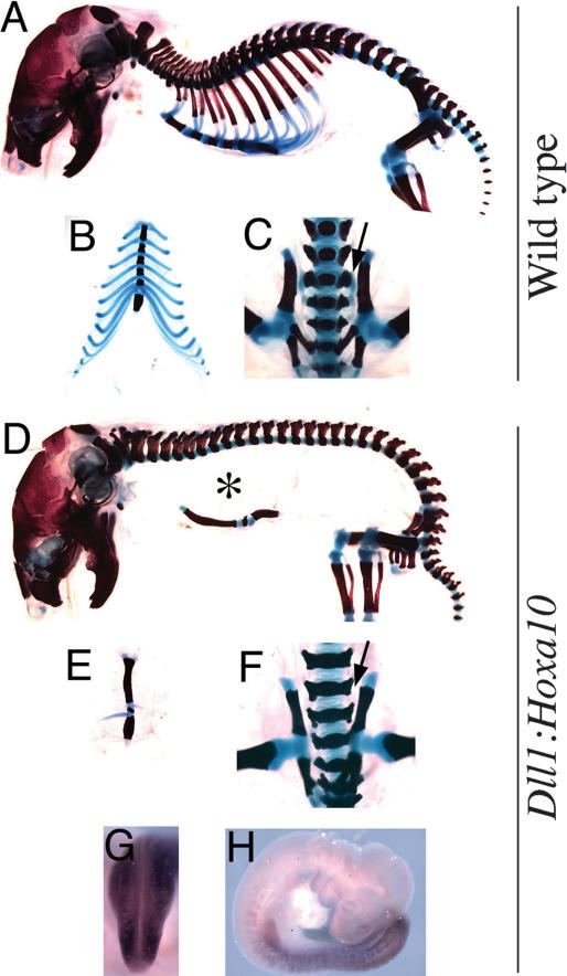
We show here that expression of Hoxa10 in the presomitic mesoderm is sufficient to confer a Hox group 10 patterning program to the somite, producing vertebrae without ribs, an effect not achieved when Hoxa10 is expressed in the somites. In addition, Hox group 11-dependent vertebral sacralization requires Hoxa11 expression in the presomitic mesoderm, while their caudal differentiation requires that Hoxa11 is expressed in the somites. Therefore, Hox gene patterning activity is different in the somites and presomitic mesoderm, the latter being very prominent for Hox gene-mediated patterning of the axial skeleton. This is further supported by our finding that inactivation of Gbx2, a homeobox-containing gene expressed in the presomitic mesoderm but not in the somites, produced Hox-like phenotypes in the axial skeleton without affecting Hox gene expression.
Figures
 indicates the position of vertebra 20, which is T13 in wild-type embryos; the
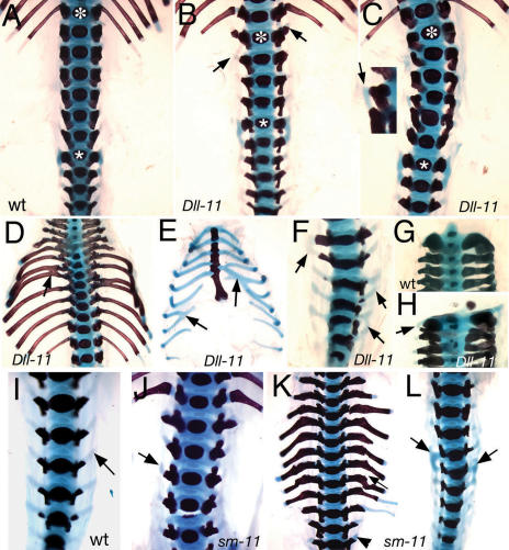
  indicates S1. (B,C) Ventral views of two Dll1-Hoxa11 transgenic embryos. In the embryo in B, S1 () is located in vertebra 24, and contains lateral protrusions in lumbar vertebrae (arrow). In the embryo in C, S1 is vertebra 26 and contains a lateral fusion between adjacent lumbar vertebrae (arrow in the blown-up region). (D) Thoracic area of a Dll1-Hoxa11 transgenic newborn showing fusions between the ossified area of adjacent ribs (arrows). (E) Sternum and the cartilaginous area of the ribcage of a Dll1-Hoxa11 transgenic newborn showing fusions between adjacent ribs (arrows). (F) Caudal region of a Dll1-Hoxa11 transgenic newborn showing fusions between adjacent vertebrae (arrows). (G,H) Cervical area of a wild-type (G) and a Dll1-Hoxa11 transgenic (H) newborn showing a lateral cartilaginous fusion between adjacent vertebrae (arrow in H). (I) Caudal area of a wild-type embryo. The arrow indicates the lateral process in a caudal vertebra. (J-L) The upper lumbar (J), thoracic (K), and sacral (L) areas of specific sm-Hoxa11 transgenic newborns. The arrow in J indicates an anteriorly projecting protuberance in a lumbar vertebra. The arrow in K indicates an anteriorly projecting protuberance at the base of a rib, and the arrowhead indicates an anteriorly projecting protuberance in a lumbar vertebra. The arrows in L show the unilateral anteriorization of the sacrum.
References
-
- Beckers J., Caron, A., Hrabe de Angelis, M., Hans, S., Campos-Ortega, J.A., and Gossler, A. 2000. Distinct regulatory elements direct δ1 expression in the nervous system and paraxial mesoderm of transgenic mice. Mech. Dev. 95: 23-34. - PubMed
-
- Bobola N., Carapuco, M., Ohnemus, S., Kanzler, B., Leibbrandt, A., Neubuser, A., Drouin, J., and Mallo, M. 2003. Mesenchymal patterning by Hoxa2 requires blocking Fgf-dependent activation of Ptx1. Development 130: 3403-3414. - PubMed
-
- Bouillet P., Chazaud, C., Oulad-Abdelghani, M., Dolle, P., and Chambon, P. 1995. Sequence and expression pattern of the Stra7 (Gbx-2) homeobox-containing gene induced by retinoic acid in P19 embryonal carcinoma cells. Dev. Dyn. 204: 372-382. - PubMed
-
- Burke A.C., Nelson, C.E., Morgan, B.A., and Tabin, C. 1995. Hox genes and the evolution of vertebrate axial morphology. Development 121: 333-346. - PubMed
-
- Chawengsaksophak K., James, R., Hammond, V.E., Kontgen, F., and Beck, F. 1997. Homeosis and intestinal tumours in Cdx2 mutant mice. Nature 386: 84-87. - PubMed
Publication types
MeSH terms
Substances
LinkOut - more resources
Full Text Sources
Molecular Biology Databases
Research Materials