. 2005 Sep 15;19(18):2236-44.
doi: 10.1101/gad.1335705.

Evidence that entry into sporulation in Bacillus subtilis is governed by a gradual increase in the level and activity of the master regulator Spo0A

Affiliations

Evidence that entry into sporulation in Bacillus subtilis is governed by a gradual increase in the level and activity of the master regulator Spo0A

Masaya Fujita et al. Genes Dev. .

Abstract

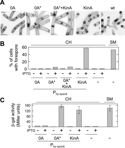
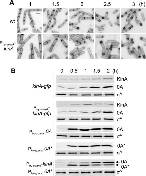
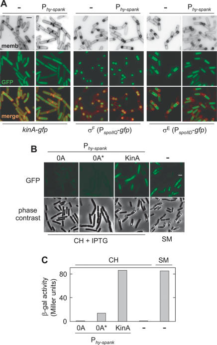
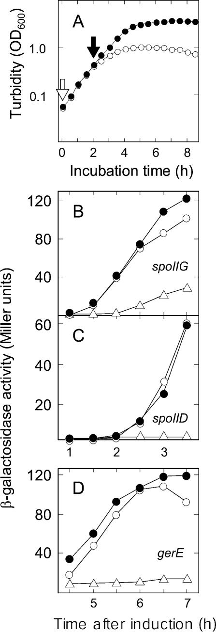
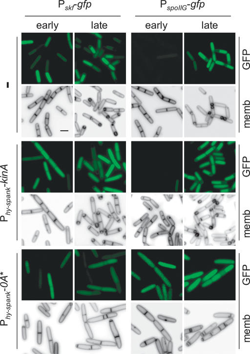
The transcription factor Spo0A is a master regulator for entry into sporulation in the bacterium Bacillus subtilis, but it has been uncertain whether activation of Spo0A is sufficient to trigger development. Spo0A, a member of the response regulator family of gene-control proteins, is activated by phosphorylation via a multicomponent phosphorelay in response to conditions of nutrient limitation. We now report that sporulation can be triggered with high efficiency in cells in the exponential phase of growth in rich medium by artificial induction of the synthesis of any one of three histidine kinases that feed phosphoryl groups into the relay. We further show that the levels of Spo0A protein and activity increase gradually over the first 2 h of sporulation both under conditions of nutrient limitation and in response to induction of kinase synthesis. Evidence indicates that this gradual increase in Spo0A protein and activity plays a critical role in triggering sporulation and requires the action of the phosphorelay.


Figures

Figure 1.
Figure 1.
Triggering efficient sporulation by artificial induction of KinA synthesis. (A) Forespore formation. Cells of strains MF2001 (Phy-spank-spo0A), MF2146 (Phy-spank-spo0A*), MF2252 (Phy-spank-kinA, Phy-spank-spo0A*), and MF1887 (Phy-spank-kinA) were grown in CH medium and treated with IPTG (20 μM for MF1887, and 200 μM for MF2001, MF2146, and MF2252) at the mid-exponential phase of growth. Cells of the wild-type strain PY79 (wt) were suspended in SM sporulation medium. Cells were treated with the vital membrane stain FM4-64 at hour 3 after suspension in SM medium for strain PY79 (wt) or after treatment with inducer for the strains harboring IPTG-inducible constructs, and observed by fluorescence microscopy. IPTG was used at concentrations determined in immunoblot experiments to yield levels of KinA and Spo0A that corresponded to that observed in wild-type cells undergoing sporulation in response to nutrient limitation. Bar, 2 μm. (B) Analysis of sporulation. The percentage of sporangia with a forespore, as judged from FM4-64 staining as in A, was determined for strains engineered to produce the indicated proteins at hour 3 after the addition of inducer. Also shown as controls are the results with the wild-type strain PY79 (right-most three bars). (C) Transcription from a Spo0A-dependent promoter. The accumulation of β-galactosidase from a PspoIIG-lacZ fusion was monitored as a measure of Spo0A activity. Cells of strains MF2175 (Phy-spank-spo0A), MF2176 (Phy-spank-spo0A*), MF1917 (Phy-spank-kinA), and MF290 (-), which each contained PspoIIG-lacZ, were induced to sporulate as described in A. β-Galactosidase activity was measured at hour 2.5 after the addition of inducer for the strains harboring IPTG-inducible constructs or after suspension in SM medium for strain MF290. The average of three independent results is shown, with the error bars indicating the standard deviation.
Figure 2.
Figure 2.
Comparative timing of sporulation in response to nutrient limitation and induced synthesis of KinA. (A) Sporulation was induced in response to nutrient limitation by cells of the wild-type strain PY79 in SM medium (upper panels) or in response to KinA synthesis by cells of strain MF1887 (Phy-spank-kinA) in CH medium (lower panels). Samples were collected at the indicated times following suspension in SM medium (upper panels) or after the addition of IPTG (lower panels). The cells were treated with the membrane stain FM4-64 and examined by fluorescence microscopy. Bar, 2 μm. (B) Accumulation of KinA and Spo0A in cells of a strain (MF928; kinA-gfp) in which KinA was tagged with GFP and produced under its normal promoter and a strain (MF1996; Phy-spank-kinA-gfp) in which GFP-tagged KinA was produced under the control of an IPTG-inducible promoter. Also shown are the accumulation of Spo0A and Spo0A* in the IPTG-inducible strains MF2001 (Phy-spank-spo0A), MF2146 (Phy-spank-spo0A*), and MF2252 (Phy-spank-kinA, Phy-spank-spo0A*). Samples were collected at the indicated times after suspension in SM medium for the wild-type strain or after the addition of IPTG for all other strains and analyzed by immunoblotting using anti-GFP antibodies for KinA-GFP, and with anti-Spo0A antibodies for Spo0A and Spo0A*. The anti-σA immunoblot served as a loading control.
Figure 3.
Figure 3.
Compartment-specific gene expression in response to the induction of KinA synthesis. (A) Accumulation of KinA-GFP and compartment-specific synthesis of GFP produced under the control of σF (PspoIIQ-gfp) and σE (PspoIID-gfp) during sporulation. Cells of strains MF1996 (Phy-spank-kinA-gfp), MF1956 (Phy-spank-kinA, PspoIIQ-gfp), and MF1957 (Phy-spank-kinA, PspoIID-gfp) were induced to sporulate by addition of IPTG in CH medium. Cells of strains MF928 (-, kinA-gfp), PE128 (-, PspoIIQ-gfp), and MF248 (-, PspoIID-gfp) were induced to sporulate in SM medium as controls. The production of GFP was monitored by fluorescence microscopy at hour 1.5 for KinA-GFP and at hour 3 for PspoIIQ-gfp and PspoIID-gfp after induction of sporulation. The cells were also visualized by staining with FM4-64. Bar, 2 μm. (B) Compartment-specific synthesis of GFP produced under the control of the mother-cell regulatory protein σK. Cells of strains MF2238 (Phy-spank-spo0A, PgerE-gfp), MF2239 (Phy-spank-spo0A*, PgerE-gfp), and MF2035 (Phy-spank-kinA, PgerE-gfp) were induced to sporulate by addition of IPTG in CH medium. Cells of strain EG180 (-, PgerE-gfp) were induced to sporulate in SM medium as a control. The production of GFP was monitored by fluorescence microscopy at hour 6 after induction of sporulation. The cells were also observed by phase-contrast microscopy to detect phase-bright forespores. Bar, 2 μm. (C) Accumulation of β-galactosidase in cells harboring a fusion (PgerE-lacZ) of lacZ to a promoter under the control of σK. Cells of strain MF2243 (Phy-spank-spo0A), MF2244 (Phy-spank-spo0A*), MF2242 (Phy-spank-kinA), and MF2240 (-), which each contained PgerE-lacZ, were induced to sporulate as described in B. β-Galactosidase activity was measured at hour 6 after induction of sporulation.
Figure 4.
Figure 4.
Induction of KinA synthesis triggers sporulation early in the exponential phase of growth. (A) Effect of inducing KinA synthesis on the growth rate of cells of strain MF1913 (Phy-spank-kinA) growing in CH medium. IPTG was added at OD600 = 0.05 (open arrow and open circles) and 0.5 (filled arrow and filled circles). Cell growth was followed by turbidity measurements (OD600). (B-D) Accumulation of β-galactosidase was measured in strains harboring lacZ fused to the indicated promoters in kinA-inducible strains. Samples were collected at the indicated times after the addition of IPTG. Culture conditions and symbols are as in A. The fusions of lacZ were to the promoters for the following genes: spoIIG (B), which is under the control of Spo0A (strain MF1917); spoIID (C), which is under the control of the intermediate-stage regulatory protein σE (strain MF2210); and gerE (D), which is under the control of the late-stage regulatory protein σK (strain MF2242). Accumulation of β-galactosidase in the absence of inducer in each strain was also examined (open triangles).
Figure 5.
Figure 5.
Relative timing of transcription from low- and high-threshold promoters as monitored by use of gfp fusions. Shown are fluorescence images obtained with cells of wild-type, KinA-inducible, and Spo0A-inducible strains that each contained a fusion of gfp to the low-threshold promoter Pskf or to the high-threshold promoter PspoIIG. Cells were induced to sporulate in SM medium (in the case of the wild type) or by IPTG addition in CH medium for the strains harboring IPTG-inducible constructs. The production of GFP was monitored by fluorescence microscopy at hour 1.5 (early) and at hour 3 (late) after induction of sporulation. Strains: EG297 (-, Pskf-gfp), MF237 (-, PspoIIG-gfp), MF1959 (Phy-spank-kinA, Pskf-gfp), MF1929 (Phy-spank-kinA, PspoIIG-gfp), MF2202 (Phy-spank-spo0A*, Pskf-gfp), MF2177 (Phy-spank-spo0A*, PspoIIG-gfp). The cells were also visualized by treatment with the vital membrane stain FM4-64. Bar, 2 μm.
Figure 6.
Figure 6.
Induced synthesis of KinA, KinB, or KinC, but not of KinD or KinE, triggers sporulation. Cells of strain MF1887 (A, Phy-spank-kinA), MF1888 (B, Phy-spank-kinB), MF1889 (C, Phy-spank-kinC), MF2147 (D, Phy-spank-kinD), MF2148 (E, Phy-spank-kinE), and PY79 (wt) were induced to sporulate in CH medium followed by IPTG addition or in SM medium [PY79 (wt)]. Cells were treated with the vital membrane stain FM4-64 at hour 3 post-induction and observed by fluorescence microscopy. Bar, 2 μm.

References

    1. Ben-Yehuda S., Rudner, D.Z., and Losick, R. 2003. RacA, a bacterial protein that anchors chromosomes to the cell poles. Science 299: 532-536. - PubMed
    1. Branda S.S., Gonzalez-Pastor, J.E., Ben-Yehuda, S., Losick, R., and Kolter, R. 2001. Fruiting body formation by Bacillus subtilis. Proc. Natl. Acad. Sci. 98: 11621-11626. - PMC - PubMed
    1. Britton R.A., Eichenberger, P., Gonzalez-Pastor, J.E., Fawcett, P., Monson, R., Losick, R., and Grossman, A.D. 2002. Genome-wide analysis of the stationary-phase σ factor (σ-H) regulon of Bacillus subtilis. J. Bacteriol. 184: 4881-4890. - PMC - PubMed
    1. Burbulys D., Trach, K.A., and Hoch, J.A. 1991. Initiation of sporulation in B. subtilis is controlled by a multicomponent phosphorelay. Cell 64: 545-552. - PubMed
    1. Burkholder W.F., Kurtser, I., and Grossman, A.D. 2001. Replication initiation proteins regulate a developmental check-point in Bacillus subtilis. Cell 104: 269-279. - PubMed

Publication types

MeSH terms

LinkOut - more resources