. 2005 Oct 14;97(8):813-20.
doi: 10.1161/01.RES.0000186194.06514.b0. Epub 2005 Sep 15.

Noonan syndrome mutation Q79R in Shp2 increases proliferation of valve primordia mesenchymal cells via extracellular signal-regulated kinase 1/2 signaling

Affiliations

Noonan syndrome mutation Q79R in Shp2 increases proliferation of valve primordia mesenchymal cells via extracellular signal-regulated kinase 1/2 signaling

Maike Krenz et al. Circ Res. .

Abstract

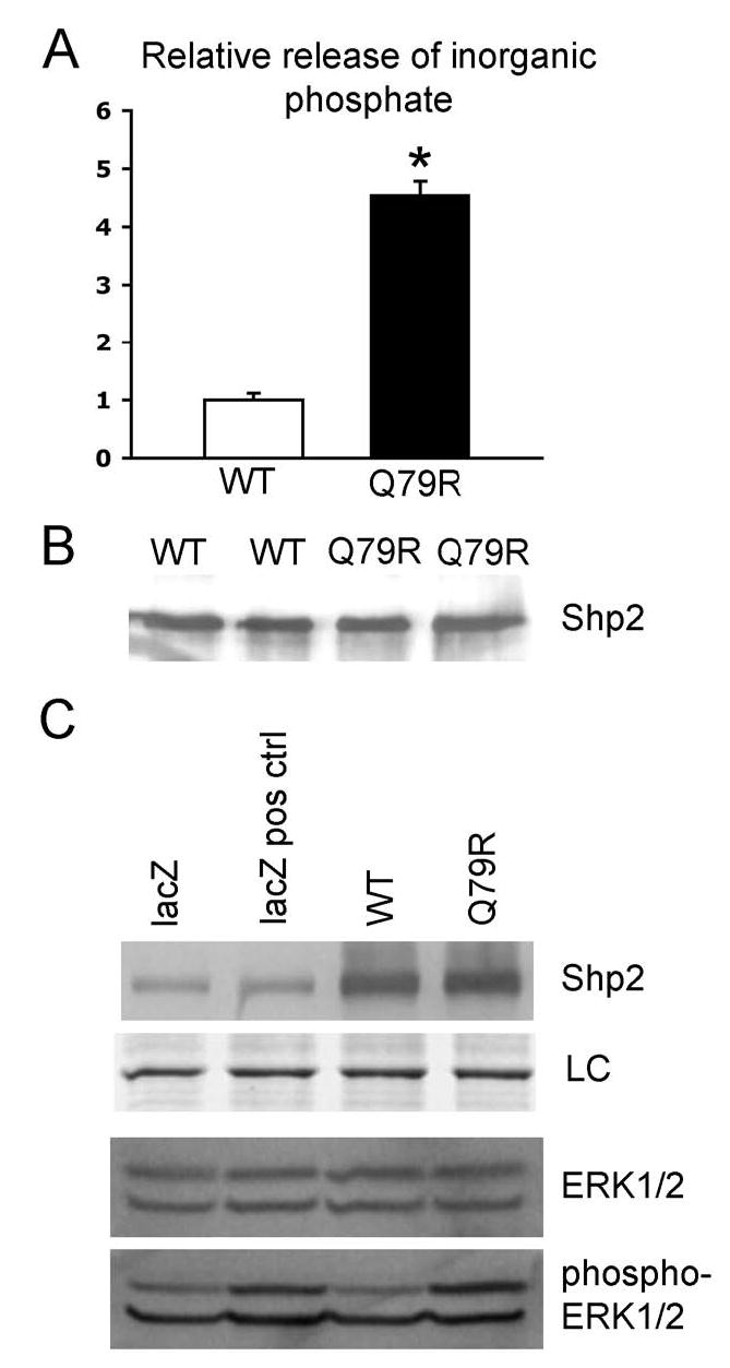
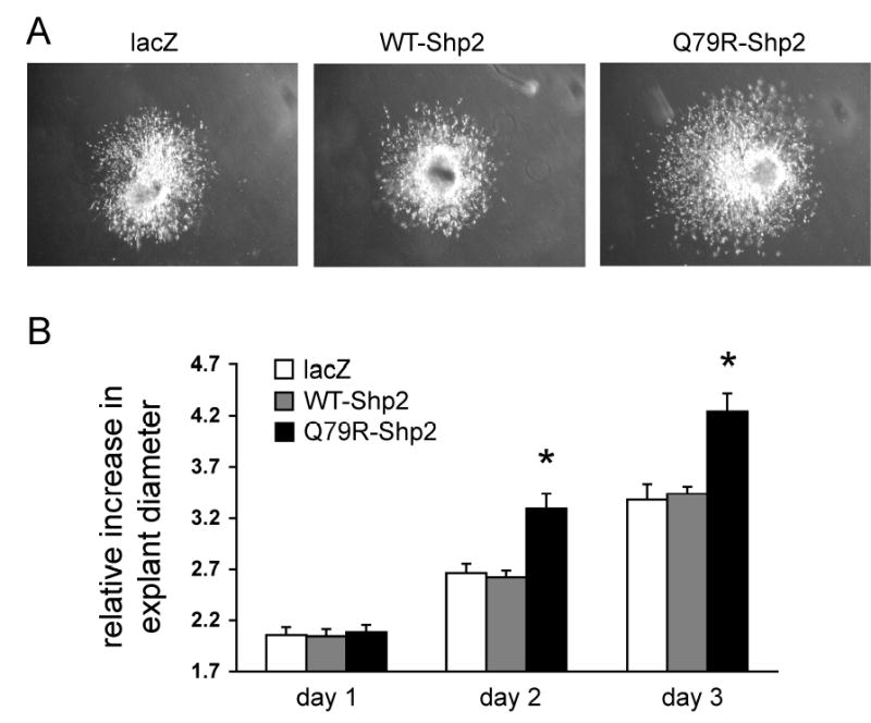
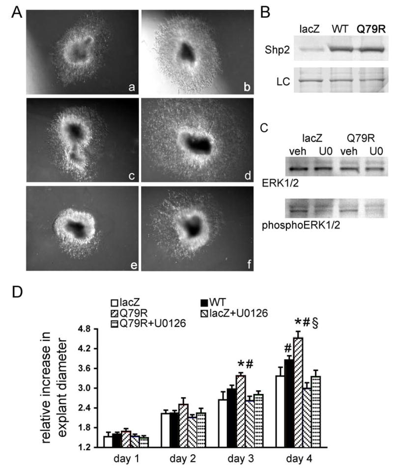
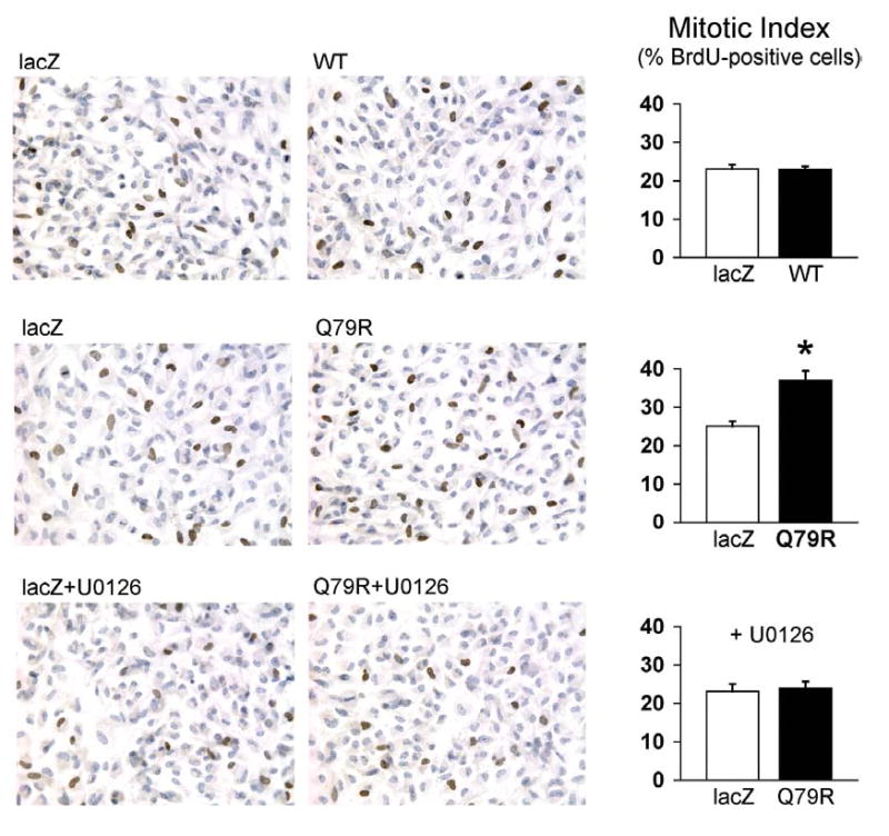
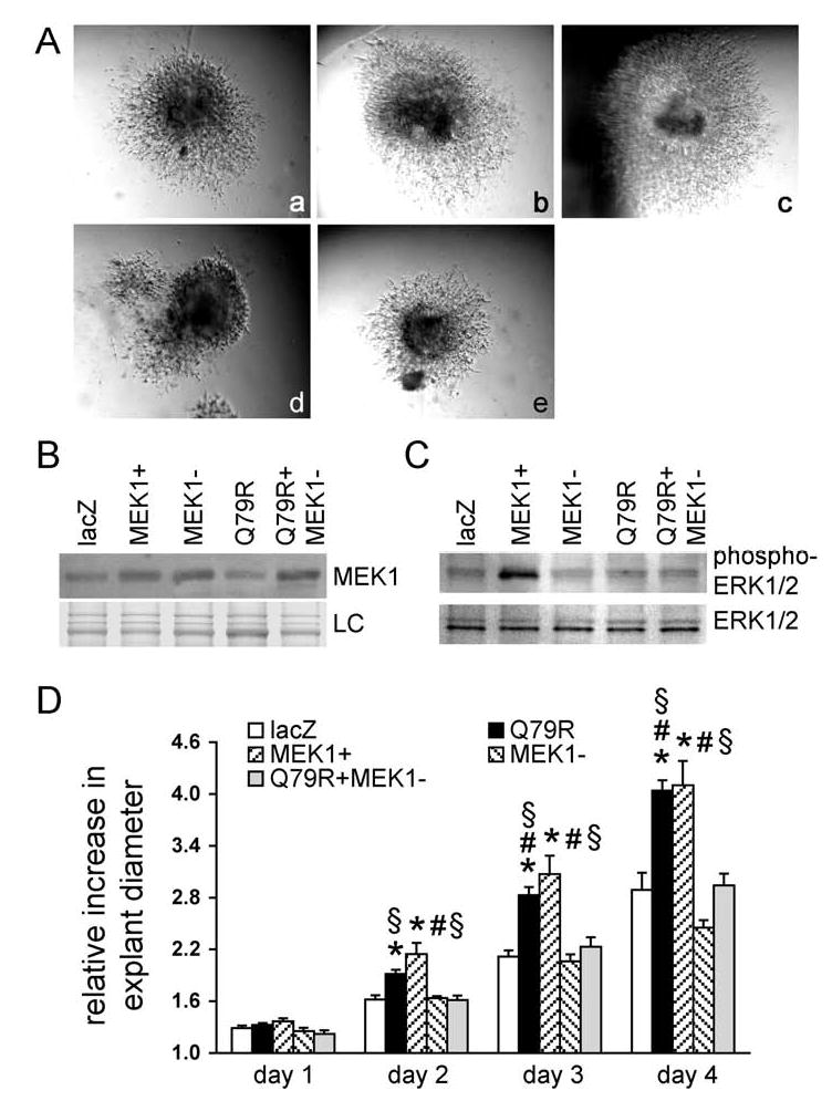
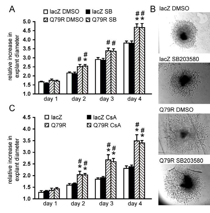
The molecular pathways regulating valve development are only partially understood. Recent studies indicate that dysregulation of mitogen-activated protein kinase (MAPK) signaling might play a major role in the pathogenesis of congenital valvular malformations, and, in this study, we explored the role of extracellular signal-regulated kinase (ERK) 1/2 activation in valve primordia expressing the Noonan syndrome mutation Q79R-Shp2. Noonan syndrome is an autosomal dominant disease characterized by dysmorphic features and cardiac abnormalities, with frequent pulmonic stenosis. The Q79R mutation of PTPN11 previously identified in Noonan syndrome families results in a gain-of-function of the encoded protein tyrosine phosphatase Shp2. We compared the effects of wild-type Shp2 and Q79R-Shp2 on endocardial cushion development. Atrioventricular and outflow tract endocardial cushions were excised from chick embryos, infected with wild-type Shp2 or Q79R-Shp2 adenovirus and embedded in a gel matrix. Q79R-Shp2, but not wild-type-Shp2, expression resulted in increased outgrowth of cells into the gel. The dependence of the Q79R-Shp2 effect on ERK1/2 and p38 MAPK signaling was then determined. The MAPK/ERK kinase (MEK)-1 inhibitor U0126, but not the p38-MAPK pathway inhibitor SB203580, abolished the effect of Q79R-Shp2 on cushion outgrowth. Coinfection with Q79R-Shp2 and dominant negative MEK-1 prevented enhanced endocardial cushion outgrowth, whereas expression of constitutively active MEK-1 mimicked the effect of Q79R-Shp2. Furthermore, dissociated cushion cells displayed increased 5-bromodeoxyuridine incorporation when infected with Q79R-Shp2 but not with wild-type Shp2. This promitotic effect was eliminated by U0126. Our results demonstrate that ERK1/2 activation is both necessary and sufficient to mediate the hyperproliferative effect of a gain-of-function mutation of Shp2 on mesenchymal cells in valve primordia.


Conflict of interest statement

Conflict-of-Interest Disclosure None.

Figures

Figure 1
Figure 1
Functional effects of the Q79R-mutation on Shp2 activity. A, Phosphatase assays with wild type and Q79R-Shp2 protein extracted from adenovirus-infected cardiomyocytes via immunoprecipitation. B, Representative immunoblot of immunoprecipitates used for phosphatase assays shown in panel A confirm that approximately equal amounts of protein were present. C, Immunoblots for Shp2 expression and ERK1/2 activation in cardiomyocytes 48 hours after adenovirus infection. As positive control, cells were stimulated with 2% fetal bovine serum for 30 min. LC, loading control (Coomassie stain of 65 – 75 kDa protein bands).
Figure 2
Figure 2
Growth rates of distal outflow tract cushion explants (Hamburger-Hamilton stage 27–28) infected with either the Q79R-Shp2 or wild type Shp2 adenoviruses. A, Representative photographs of individual explants after 3 days of culture. B, Diameters of cushion explants in culture, normalized to respective explant diameter immediately after plating. n = 10 individual explants per group, *P<0.05 versus lacZ.
Figure 3
Figure 3
Growth rates of AV cushion explants (Hamburger-Hamilton stage 25–26) infected with adenoviruses and in the presence or absence of pharmacological ERK1/2 inhibition. A, Representative photographs of individual explants after 4 days of culture. (a) lacZ, (b) WT-Shp2, (c) lacZ + DMSO, (d) Q79R-Shp2 + DMSO, (e) lacZ + U026, (f) Q79R-Shp2 + U0126. B, Representative immunoblot for Shp2 expression in AV explants after 5 days of culture. LC, loading control (Coomassie stain of 65 – 75 kDa protein bands). C, Immunoblots for ERK1/2 activation in AV explants cultured in the presence or absence of the ERK1/2 inhibitor U0126. veh, vehicle (DMSO). D, Diameters of cushion explants in culture, normalized to respective explant diameter immediately after plating, n = 4–8 per group, *P<0.05 versus lacZ, #P<0.05 versus lacZ-U0126, §P<0.05 versus QR-U0126.
Figure 4
Figure 4
BrdU-labeling of adenovirus-infected dissociated AV cushion cells grown on collagen-coated glass. Left panels, immunohistochemistry. Brown, BrdU-positive nuclei, blue, hematoxylin counterstain. Right panels, quantitative data. Counts of >2000 cells per independent experiment, n = 4 in all groups
Figure 5
Figure 5
Growth rates of AV cushion explants (Hamburger-Hamilton stage 25–26) infected with Q79R-Shp2 and MEK-1 adenoviruses. A, Representative photographs of individual explants after 4 days of culture. (a) lacZ, (b) Q79R-Shp2, (c) constitutively active MEK-1, (d) dominant negative MEK-1, (e) Q79R-Shp2 + dominant negative MEK-1. B, Representative immunoblot for MEK-1 expression in AV explants after 5 days of culture. LC, loading control (Coomassie stain of 65 – 75 kDa protein bands). C, Immunoblots for ERK1/2 activation in adenovirus-infected AV explants. D, Diameters of cushion explants in culture, normalized to respective explant diameter immediately after plating, n = 6–10 per group, *P<0.05 versus lacZ, #P<0.05 versus MEK-, §P<0.05 vs QR-MEK-.
Figure 6
Figure 6
Growth rates of AV cushion explants (Hamburger-Hamilton stage 25–26) infected with adenoviruses and in the presence or absence of p38 MAPK or calcineurin inhibition. A, Diameters of cushion explants treated with SB203580 in culture, normalized to respective explant diameter immediately after plating, n = 10–12 per group. B, Representative photographs of individual explants after 4 days of culture. B, C, Diameters of cushion explants treated with cyclosporine A (CsA) in culture, normalized to respective explant diameter immediately after plating, n = 5–6 per group. *P<0.05 vs lacZ, #P<0.05 vs lacZ + respective inhibitor.

Similar articles

Cited by

References

    1. Armstrong EJ, Bischoff J. Heart valve development: endothelial cell signaling and differentiation. Circ Res. 2004;95:459–470. - PMC - PubMed
    1. Liberatore CM, Yutzey KE. MAP kinase activation in avian cardiovascular development. Dev Dyn. 2004;230:773–780. - PubMed
    1. Allanson JE. Noonan syndrome. J Med Genet. 1987;24:9–13. - PMC - PubMed
    1. Noonan JA. Hypertelorism with Turner phenotype. A new syndrome with associated congenital heart disease. Am J Dis Child. 1968;116:373–380. - PubMed
    1. Tartaglia M, Mehler EL, Goldberg R, Zampino G, Brunner HG, Kremer H, van der Burgt I, Crosby AH, Ion A, Jeffery S, Kalidas K, Patton MA, Kucherlapati RS, Gelb BD. Mutations in PTPN11, encoding the protein tyrosine phosphatase SHP-2, cause Noonan syndrome. Nat Genet. 2001;29:465–468. - PubMed

Publication types

MeSH terms

Substances