. 2005 Oct;36(10):2258-64.
doi: 10.1161/01.STR.0000181082.70763.22. Epub 2005 Sep 15.

Neuroprotective properties of the natural vitamin E alpha-tocotrienol

Affiliations

Neuroprotective properties of the natural vitamin E alpha-tocotrienol

Savita Khanna et al. Stroke. 2005 Oct.

Abstract

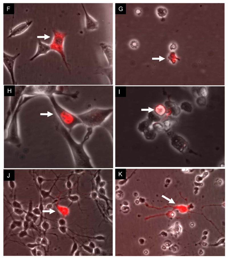
Background and purpose: The current work is based on our previous finding that in neuronal cells, nmol/L concentrations of alpha-tocotrienol (TCT), but not alpha-tocopherol (TCP), blocked glutamate-induced death by suppressing early activation of c-Src kinase and 12-lipoxygenase.

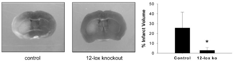
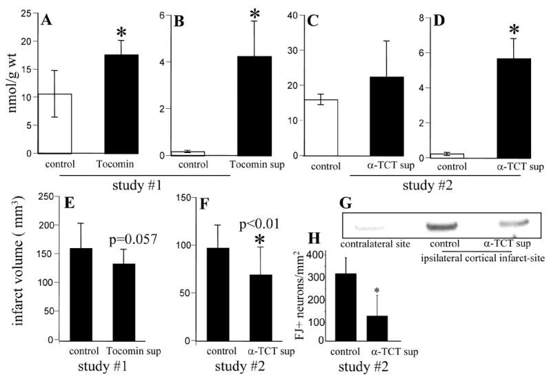
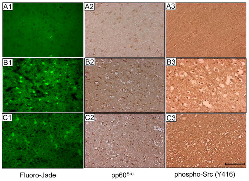
Methods: The single neuron microinjection technique was used to compare the neuroprotective effects of TCT with that of the more widely known TCP. Stroke-dependent brain tissue damage was studied in 12-Lox-deficient mice and spontaneously hypertensive rats orally supplemented with TCT.

Results: Subattomole quantity of TCT, but not TCP, protected neurons from glutamate challenge. Pharmacological as well as genetic approaches revealed that 12-Lox is rapidly tyrosine phosphorylated in the glutamate-challenged neuron and that this phosphorylation is catalyzed by c-Src. 12-Lox-deficient mice were more resistant to stroke-induced brain injury than their wild-type controls. Oral supplementation of TCT to spontaneously hypertensive rats led to increased TCT levels in the brain. TCT-supplemented rats showed more protection against stroke-induced injury compared with matched controls. Such protection was associated with lower c-Src activation and 12-Lox phosphorylation at the stroke site.

Conclusions: The natural vitamin E, TCT, acts on key molecular checkpoints to protect against glutamate- and stroke-induced neurodegeneration.


Figures

Figure 1
Figure 1. Cytosolic α-tocotrienol, but not α-tocopherol, protects neurons from glutamate induced death
HT4 (A–I) were injected with α-tocotrienol (10−19 mole) into the cytoplasm (A; 90% survival count in six experiments) or nucleus (D). Panel B shows survival of the neuron injected with α-tocotrienol while the other neurons died and disappeared from the monolayer. Tocotrienol was co-injected with the fluorescent QDot (see in red). The culture plate containing the surviving cell was repopulated with fresh healthy HT4 cells to monitor the fate the surviving cell (arrow marked in C) over a period of 18h. Calcein AM was used to stain live cells (C). Control cells injected with QDot alone (not shown) or with tocopherol (Fig. F–G, cytosolic; H–I, nuclear) did not survive (0% survival count in five experiments) against glutamate-induced challenge. Cytosolic injection of α-tocotrienol protected primary immature cortical neurons (J–K) against glutamate challenge as well. All panels in the left column represent images at 0h of glutamate challenge. All panels in the right column represent images at 18h of 10 mM glutamate challenge. Representative illustrations of five experiments are shown. Objectively, nuclear injection of α-tocotrienol failed to protect in 100% case. α-Tocopherol (10−19 moles) failed to protect in 100% case. Cytosolic injection of α-tocotrienol protected cells in 90% of all cases. Arrows in each pair of photos (left-right)
Figure 2
Figure 2. c-Src and 12-Lox in glutamate-induced neuronal death
HT4 cells (A–B) were either treated or not with α-tocotrienol, BL15, herbimycin or geldanamycin (as indicated) for 5 min and challenged with buthionine sulfoximine (0.15 mM; BSO, A) or BSO and arachidonic acid (0.05 mM, B) for 24 h. BL 15 is an inhibitor of 12-lipoxygenase. Both herbimycin and geldanamycin inhibit c-Src kinase activity. A, Tocotrienol, 12-Lox inhibitor as well as c-Src inhibitors protected against BSO-induced glutathione-depletion dependent loss of cell viability. †, higher than BSO non-treated; *, lower than BSO-treated. B, Tocotrienol, 12-Lox inhibitor, and c-Src kinase inhibitors protected against BSO and arachidonic acid induced loss of cell viability. †, higher than BSO (A) and non-challenged (B) groups; *, lower than BSO and arachidonic acid challenged group. p < 0.001. Glutamate (10 mM) induced 12-Lox phosphorylation was inhibited by α-tocotrienol (250 nmol/L) in HT4 cells (C) as well as in immature cortical neurons (F). Herbimycin also inhibited inducible 12-Lox phosphorylation (E). Cells were either treated or not with α-tocotrienol for 5 min and challenged with glutamate (10 mM) or BSO (0.15 mM) and arachidonic acid (0.05 mM) for 15 min (D) or 30 min as indicated. In these experiments 15 minutes before challenging, cells were treated with Na3VO4 (0.15 mM) to inhibit tyrosine phosphatases. C, control (non-treated); TCT, α-tocotrienol; H, herbimycin; G, glutamate; and AA, arachidonic acid. G–H, Glutamate-induced phosphorylation was inhibited (G) in cells expressing dominant negative c-Src (K296R/Y528F) and more prominent in kinase-active (Y529F) c-Src over-expressing cells than in wild-type (pUSE) or kinase-dead c-Src (K297R) over-expressing cells (H). Cells were activated with 10 mM glutamate for 15 mins.
Figure 3
Figure 3. 12-Lipoxygenase is phosphorylated by c-Src and phospho-12-lipoxygenase activity is sensitive to α-tocotrienol
In the presence of ATP, c-Src phosphorylated 12-Lox in a herbimycin (A) and PP2 (B) sensitive manner. PP3, a negative control for PP2, did not inhibit 12-Lox phosphorylation indicating a specific role of c-Src. C, α-Tocotrienol inhibited the activity of 12-Lox as well as of phospho-12-Lox. Phospho-12-Lox was generated (as in A and B) by incubation of the enzyme with c-Src and ATP. †, p<0.05 significant increase in conjugated diene (reaction product) formation in response to 12-Lox addition to the reaction mix. *, p<0.05 lower activity of both 12-Lox as well as phospho-12-Lox in the presence of α-tocotrienol (TCT) compared to the activity observed in the absence of TCT. Values are mean ± SD.
Figure 4
Figure 4. 12-Lipoxygenase deficient mice are resistant to transient focal cerebral ischemia
Transient focal cerebral ischemia was induced in the C57BL6/J (control) and 12-lipoxygenase knockout (12-Lox −/−) mice by middle cerebral artery occlusion. Coronal sections of brain (72h after stroke) were stained with 2,3,5-triphenyltetrazolium (TTC). Fixed sections were photographed using a Inquiry software (Loats Associates, Inc, Westminster, MD). The images were used to determine infarct size as a percentage of the contralateral hemisphere after correcting for edema. The infarct extended from caudate putamen into surrounding cortex, and was visible in 4 out of 5 slices of the brain from control wild-type mice. Data are mean ± SD. *, p<0.02.
Figure 5
Figure 5. Oral α-tocotrienol supplementation protected against stroke induced injury and 12-lipoxygenase phosphorylation in the brain of spontaneously hypertensive rats
The effect of oral supplementation on brain α tocopherol (A,C) and α tocotrienol (B,D). *, p<0.01 significantly different compared to the corresponding control group. Infarct volume (E,F) in response to stroke. G, α-Tocotrienol supplementation lowered 12-lipoxygenase phosphorylation in the stroke-site brain tissue of SHR. The brain tissue sample was harvested 24h after stroke from a predictable area of infarct core around the occluded vessel. This includes the primary motor cortex and the primary somatosensory cortex. H, Histological analyses of post-stroke infarct zone cortical section (Figure 6) revealed lower count of Fluoro-Jade positive (FJ+) neurons in tocotrienol supplemented rats. *, p<0.05. These results represent the image analysis data of sections (A1, B1 & C1) shown in Figure 6. Study I, n=16; Study II, n=21.
Figure 6
Figure 6. Histological analyses of post-stroke infarct zone cortical sections
Fluoro-Jade (FJ) stain. In the contralateral hemisphere of the brain from control rats (A1) there was hardly any FJ-positive neuron. The ipsilateral hemisphere of the brain from α-tocotrienol supplemented rats (C1) contained fewer (see Figure 5H) FJ-positive cells compared to such cells in the ipsilateral hemisphere of the brain from control rats (B1). Src and phospho-Src staining. In non-stroke site of the brain section, Src was present (A2) but not in a phosphorylated form (A3). Stroke did not influence Src expression but clearly induced Src phosphorylation and activation. Such stroke-induced Src activation was less in the brain sections of tocotrienol-supplemented rats compared to control rats that were subjected to stroke (C3 versus B3). For all sections shown, brain was harvested 24h after stroke. Bar = 100 micron.

References

    1. Brigelius-Flohe R, Traber MG. Vitamin e: Function and metabolism. FASEB Journal. 1999;13:1145–1155. - PubMed
    1. Traber MG, Packer L. Vitamin e: Beyond antioxidant function. Am J Clin Nutr. 1995;62:1501S–1509S. - PubMed
    1. Traber MG, Sies H. Vitamin e in humans: Demand and delivery. Annu Rev Nutr. 1996;16:321–347. - PubMed
    1. Sen CK, Khanna S, Roy S. Tocotrienol: The natural vitamin e to defend the nervous system? Ann New York Acad Sci. 2004;1031:127–142. - PubMed
    1. Sen CK, Khanna S, Roy S, Packer L. Molecular basis of vitamin e action. Tocotrienol potently inhibits glutamate-induced pp60(c-src) kinase activation and death of ht4 neuronal cells. Journal of Biological Chemistry. 2000;275:13049–13055. - PubMed

Publication types

MeSH terms