Comparative Study
. 2006 Feb 15;107(4):1651-8.
doi: 10.1182/blood-2005-07-2839. Epub 2005 Sep 15.

Pathology of Berkeley sickle cell mice: similarities and differences with human sickle cell disease

Affiliations
Comparative Study

Pathology of Berkeley sickle cell mice: similarities and differences with human sickle cell disease

Elizabeth A Manci et al. Blood. .

Abstract

Because Berkeley sickle cell mice are used as an animal model for human sickle cell disease, we investigated the progression of the histopathology in these animals over 6 months and compared these findings to those published in humans with sickle cell disease. The murine study groups were composed of wild-type mixed C57Bl/6-SV129 (control) mice and sickle cell (SS) mice (alpha-/-, beta-/-, transgene +) of both sexes and between 1 and 6 months of age. SS mice were similar to humans with sickle cell disease in having erythrocytic sickling, vascular ectasia, intravascular hemolysis, exuberant hematopoiesis, cardiomegaly, glomerulosclerosis, visceral congestion, hemorrhages, multiorgan infarcts, pyknotic neurons, and progressive siderosis. Cerebral perfusion studies demonstrated increased blood-brain barrier permeability in SS mice. SS mice differed from humans with sickle cell disease in having splenomegaly, splenic hematopoiesis, more severe hepatic infarcts, less severe pulmonary manifestations, no significant vascular intimal hyperplasia, and only a trend toward vascular medial hypertrophy. Early retinal degeneration caused by a homozygous mutation (rd1) independent from that causing sickle hemoglobin was an incidental finding in some Berkeley mice. While our study reinforces the fundamental strength of this model, the notable differences warrant careful consideration when drawing parallels to human sickle cell disease.


Figures
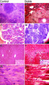
Figure 1.
Figure 1.
Comparison of histopathology in control and sickle mice. The columns show microscopic findings in control (A, C, E, G) and sickle (B, D, F, H) mice. The 4 rows (top to bottom) show findings in the spleen (A, B), bone marrow (C, D), liver (E, F), and heart (G, H). (A, B) Sickle mice had severe splenic sinusoidal congestion by sickle erythrocytes, increased hematopoietic cells, infarcts (arrows), and vascular ectasia (inset) (H&E original magnification, 100 ×; inset, 400 ×). (C, D) Erythroid hyperplasia was more severe in the bone marrow of sickle mice (H&E original magnification, 100 ×). (E, F) Hepatic findings in sickle mice included more sinusoidal congestion, infarct, and siderosis (inset) (H&E original magnification, 100 ×; insets iron stain, 400 ×). (G, H) Cardiac findings in sickle mice included infarcts, which, when acute, appeared as loss of normal striations (arrows).
Figure 2.
Figure 2.
Comparison of histopathology in control and sickle mice. The columns show microscopic findings in control (A, C, E, G) and sickle (B, D, F, H) mice. The 4 rows (top to bottom) show findings in the lung (A, B), kidney (C, D), glomerulus (E, F), and cerebellum (G, H). (A, B) Pulmonary findings in the sickle mice included more severe vascular congestion (arrows) and siderosis (inset) (H&E original magnification, 100 ×; inset iron stain, 100 ×). (C, D) Renal findings included infarcts (arrows) and siderosis (inset) in the sickle mice (Trichrome stain original magnification, 100 ×; inset iron stain, 100 ×). (E, F) Glomerular changes in the sickle mice included mesangial hyperplasia (arrows). (Trichrome stain original magnification, 400 ×). (G, H) Cerebellar findings included pyknotic Purkinje cell in the sickle mice (arrows) (Trichrome stain original magnification, 100 ×).
Figure 3.
Figure 3.
Cerebral microvessels perfused with FITC-dextran. The plasma-demarcated capillary networks show a broad array of twists, turns, and junctions in the striatum from a control mouse (A). Irregularly dilated cerebral microvessels with leakage of FITC-dextran was detected in the striatum of an SS mouse (B, C). Panel C is a high magnification of boxed area in panel B (Bar = 40 μm).
Figure 4.
Figure 4.
Retinas from 1-year-old Berkeley SS mice that were heterozygous or homozygous for the rd1 mutation. (A, C, E) Heterozygous for the rd1 mutation; (B, D, F) homozygous for the rd1 mutation. Retinas were incubated for enzyme histochemical demonstration of ADPase activity and then flat-embedded in JB-4. The ADPase activity in viable blood vessels appears white with dark-field illumination (A, B) and black with bright-field illumination (C, D). (A) The normal mouse retina has a spoke wheel pattern of blood vessels that emanates from the optic nerve head (center). (B) The Berkeley mouse that was homozygous for rd1 had a much attenuated retinal vasculature and was missing the dense, deep capillary system. (C) There was no pigment present in the retina of the mouse heterozygous for rd1 with bright-field illumination. (D) The Berkeley rd1/rd1 mouse had many areas that were pigmented, suggesting that the retinal pigment epithelial (RPE) cells had migrated into the sensory retina (arrow). (E) When the normal-appearing retina in panels A and C was sectioned, the 2 nuclear layers of retina were apparent, and photoreceptor outer segments were present below the outer nuclear layer (bottom). The retinal vasculature in the inner retina appeared normal, and a vessel is seen connecting the superficial and deep capillary systems of the retina in this field. (F) The Berkeley rd1/rd1 mouse had no outer nuclear layer or photoreceptor outer or inner segments (bottom), a disorganized inner nuclear layer (bottom), and RPE cells (arrow) had migrated into the retina and ensheathed a dilated blood vessel.

Comment in

Similar articles

Cited by

References

    1. Paszty C. Transgenic and gene knock-out mouse models of sickle cell anemia and the thalassemias. Curr Opin Hematol. 1997;4: 88-93. - PubMed
    1. Paszty C, Brion CM, Manci E, et al. Transgenic knockout mice with exclusively human sickle hemoglobin and sickle cell disease. Science. 1997; 278: 876-878. - PubMed
    1. de Jong K, Emerson RK, Butler J, Bastacky J, Mohandas N, Kuypers FA. Short survival of phosphatidylserine-exposing red blood cells in murine sickle cell anemia. Blood. 2001;98: 1577-1584. - PubMed
    1. Fabry ME, Suzuka SM, Weinberg RS, et al. Second generation knockout sickle mice: the effect of HbF. Blood. 2001;97: 410-418. - PubMed
    1. Kean LS, Manci EA, Perry J, et al. Chimerism and cure: hematologic and pathologic correction of murine sickle cell disease. Blood. 2003;102: 4582-4593. - PubMed

Publication types