. 2006 Feb 1;173(3):318-26.
doi: 10.1164/rccm.200503-425OC. Epub 2005 Sep 15.

Imbalance between cysteine proteases and inhibitors in a baboon model of bronchopulmonary dysplasia

Affiliations

Imbalance between cysteine proteases and inhibitors in a baboon model of bronchopulmonary dysplasia

Ozden Altiok et al. Am J Respir Crit Care Med. .

Abstract

Rationale: Bronchopulmonary dysplasia (BPD) continues to be a major morbidity in preterm infants. The lung pathology in BPD is characterized by impaired alveolar and capillary development. An imbalance between proteases and protease inhibitors in association with changes in lung elastic fibers has been implicated in the pathogenesis of BPD.

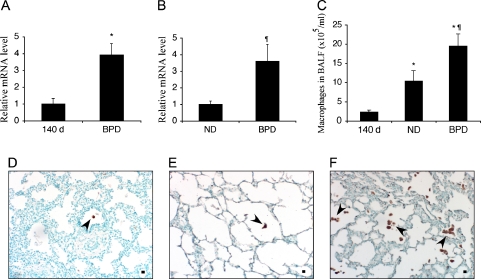
Objective: To investigate the expression and activity levels of papain-like lysosomal cysteine proteases, cathepsins B, H, K, L, S, and their inhibitors, cystatins B and C, in a baboon model of BPD.

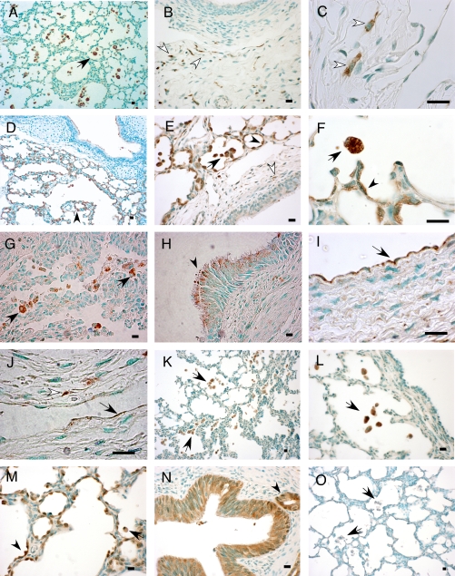
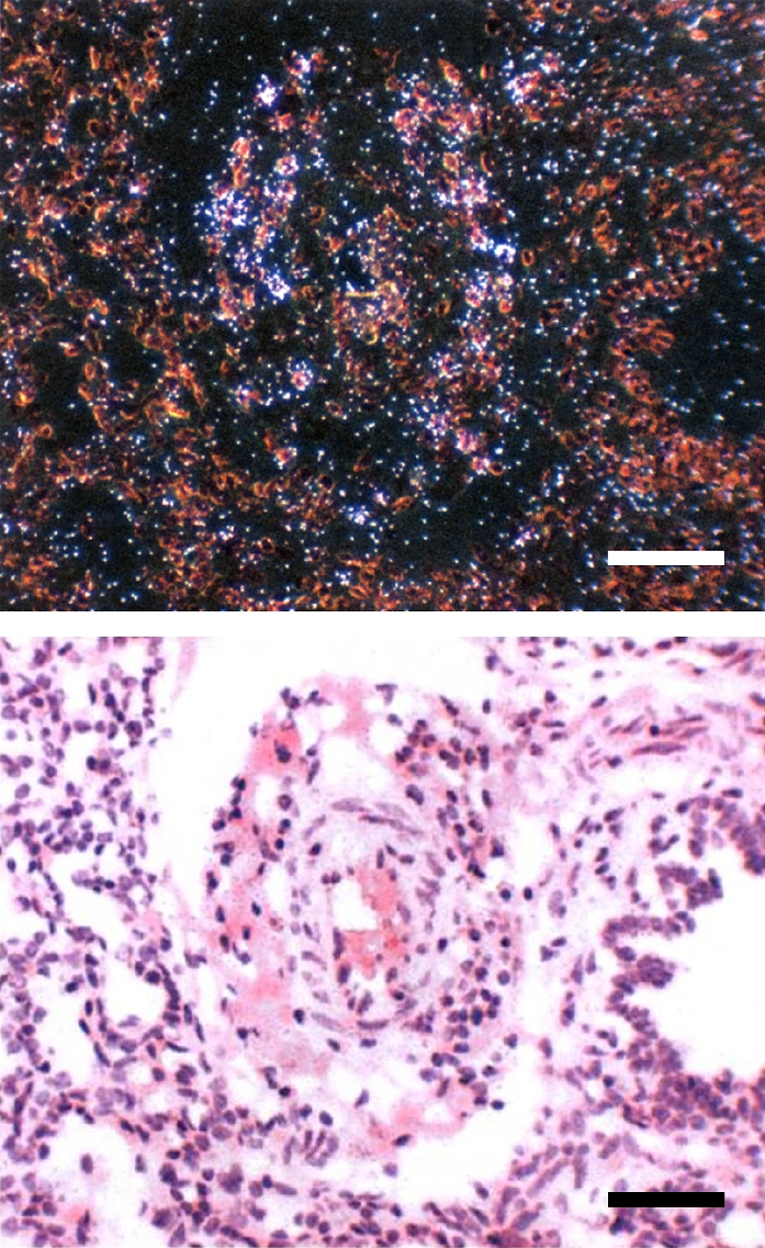
Methods: Real-time reverse transcriptase-polymerase chain reaction, immunohistochemistry, immunoblotting, active site labeling of cysteine proteases, and in situ hybridization were performed.

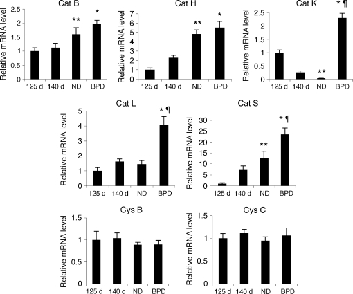
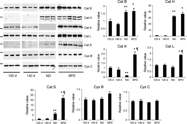
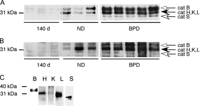
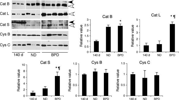
Measurements and main results: The steady-state mRNA and protein levels of all cathepsins were significantly increased in the lung tissue of baboons with BPD. In contrast, the steady-state mRNA and protein levels of two major cysteine protease inhibitors, cystatin B and C, were unchanged. Correlating with these alterations, the activity of cysteine proteases in lung tissue homogenates and bronchoalveolar lavage fluid was significantly higher in the BPD group. The levels of cathepsin B, H, and S increased and cathepsin K decreased with advancing gestation. All cathepsins, except for cat K, were immunolocalized to macrophages in BPD. In addition, cathepsin H and cystatin B were colocalized in type 2 alveolar epithelial cells. Cathepsin L was detected in some bronchial epithelial, endothelial, and interstitial cells. Cathepsin K was localized to some perivascular cells by in situ hybridization.

Conclusions: Cumulatively, these findings demonstrate an imbalance between cysteine proteases and their inhibitors in BPD.


Figures

<b>Figure 1.</b>
Figure 1.
Cathepsin mRNA levels during lung development and in BPD. Relative steady-state mRNA levels of cathepsin (cat) B, cat H, cat K, cat L, cat S, cystatin (cys) B, and cys C were determined by quantitative reverse transcriptase–polymerase chain reaction (RT-PCR) using RNA obtained from 125-d, 140-d, natural delivery (ND), and bronchopulmonary dysplasia (BPD) group baboons. n = 6–8 animals/group. Data are expressed as mean ± SEM from two experiments performed in triplicates. *p < 0.05 versus 140 d; **p < 0.05 versus 125 d; p < 0.05 versus ND.
<b>Figure 2.</b>
Figure 2.
Cathepsin (cat) proteins during lung development and in BPD. Immunoblot and densitometry analysis for cat B, H, K, L, and S, and cystatin (cys) B and C proteins were performed in total lung homogenates obtained from 125-d, 140-d, ND, and BPD group baboons. Results are representative of three independent experiments using a total of 6–10 different study animals/group. The marker indicates 28 and 15 kD for cathepsin and cystatin immunoblots, respectively. Mean ± SEM values for all proteins are normalized to values in the 125-d gestation lungs. The total amount of protein loaded per lane is 4 μg for cat L, 5 μg for cys B, 10 μg for cat B, H, and K, and 15 μg for cat S immunoblots. *p < 0.05 versus 140 d; **p < 0.05 versus 125 d; p < 0.05 versus ND.
<b>Figure 3.</b>
Figure 3.
Increased activity of cathepsins in BPD. Lung lysates (A) and bronchoalveolar lavage fluid (BALF; B) obtained from 140-d, ND, and BPD group baboons were normalized to total protein, incubated with a biotin-labeled E-64 analog (JPM-565), and resolved by sodium dodecyl sulfate–polyacrylamide gel electrophoresis and chemiluminescence. Data are representative of three independent experiments using a total of 6–10 different study animals/group. (C) To identify the relative molecular masses of active site–labeled cathepsins in the baboon lung, active site–labeled and pooled lung homogenates from three baboons in the BPD group were immunoprecipitated using cat B, H, K, L, and S antibodies. All immunoprecipitated cathepsins were detected with horseradish peroxidase–streptavidin and chemiluminescence.
<b>Figure 4.</b>
Figure 4.
Detection of cathepsins (cat) in BALF. Immunoblot analysis for cat B, L, and S, and cystatin (cys) B and C were performed on baboon necropsy lavage fluids of 140-d, ND, and BPD group baboons. Results are representative of at least two experiments using samples from a total of 6–10 different study animals/group. Black and white arrows indicate procathepsins and mature cathepsins, respectively. The total amount of protein loaded per lane is 4 μg for cat B and cys B and C, and 7 μg for cat L and S immunoblots. *p < 0.05 versus 140 d; p < 0.05 versus ND.
<b>Figure 5.</b>
Figure 5.
Macrophages in baboon lung tissues and BALF in BPD. Relative steady-state mRNA levels of the activated macrophage marker CD68 were determined by quantitative RT-PCR using RNA obtained from total baboon lung homogenates (A) or BAL cells (B); n = 4–5 animals/group. Data are expressed as mean ± SEM from two experiments performed in triplicate. Absolute macrophage numbers were determined in BAL cell pellets of 140-d, ND, and BPD group animals (C); n = 10–12 animals/group. Immunohistochemistry was performed on paraffin-embedded lung tissue sections from 140-d (D), ND (E), and BPD (F) groups using a CD68 antibody. Methyl green was used as the counterstain. Arrowheads point to CD68-positive macrophages in representative tissue sections. Scale bar, 100 μm. *p < 0.05 versus 140 d; p < 0.05 versus ND.
<b>Figure 6.</b>
Figure 6.
Immunolocalization of cathepsin and cystatin B proteins in BPD. Immunohistochemistry was performed on formalin-fixed, paraffin-embedded lung tissue sections of baboons with BPD using specific antibodies for cat B, H, L, and S, and cystatin B. Methyl green was used as the counterstain. Photomicrographs are representative of the results obtained in a minimum of four experiments using samples from four to five study animals. Scale bar, 100 μm. Shown are cat B immunoreactivity in macrophages (A; black arrow), interstitial cells (B; white arrows), and fibroblasts (C; white arrows); cat H staining in type 2 alveolar epithelial cells (D, E, and F; arrowheads), macrophages (D, E, and F; black arrows), and rare interstitial cells (E; white arrows); cat L immunoreactivity in macrophages (G; black arrows), bronchial epithelial cells in supranuclear localization (H; black arrowhead), vascular (I; black arrow) and lymphatic endothelial cells (J; black arrow), and a fibroblast (J; white arrowhead); cat S staining in macrophages (K and L; black arrows); cystatin B immunoreactivity in type 2 alveolar epithelial cells (M; arrowhead), macrophages (M; black arrow), and bronchial epithelial cells (N) and a bronchial gland (N; black arrowhead); negative control stained with mouse primary antibody isotype control showing no staining in macrophages (O; black arrows).
<b>Figure 7.</b>
Figure 7.
Cat K mRNA expression in BPD. In situ hybridization using a 35S-labeled antisense riboprobe specific for cat K was performed. Shown are paired bright and dark-field views of lung sections hybridized in situ for cat K mRNA. Positive signal is visible as white silver grains in the dark-field view. Note individual positive cells localizing to perivascular areas. Scale bar, 100 μm.

References

    1. Northway WJ, Rosan RC, Porter DY. Pulmonary disease following respirator therapy of hyaline-membrane disease: bronchopulmonary dysplasia. N Engl J Med 1967;276:357–368. - PubMed
    1. Bonikos DS, Bensch KG. Pathogenesis of bronchopulmonary dysplasia. In: Merritt TA, Northway WH, editors. Bronchopulmonary dysplasia. Boston, MA: Blackwell; 1988. pp. 33–58.
    1. Bancalari E, Claure N, Sosenko IR. Bronchopulmonary dysplasia: changes in pathogenesis, epidemiology and definition. Semin Neonatol 2003;8:63–71. - PubMed
    1. Tanswell AK, Jankov RP. Bronchopulmonary dysplasia: one disease or two? Am J Respir Crit Care Med 2003;167:1–2. - PubMed
    1. Coalson JJ. Pathology of new bronchopulmonary dysplasia. Semin Neonatol 2003;8:73–81. - PubMed

Publication types