. 2005 Oct;25(19):8581-91.
doi: 10.1128/MCB.25.19.8581-8591.2005.

The Runx2 osteogenic transcription factor regulates matrix metalloproteinase 9 in bone metastatic cancer cells and controls cell invasion

Affiliations

The Runx2 osteogenic transcription factor regulates matrix metalloproteinase 9 in bone metastatic cancer cells and controls cell invasion

Jitesh Pratap et al. Mol Cell Biol. 2005 Oct.

Abstract

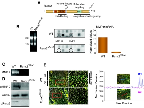
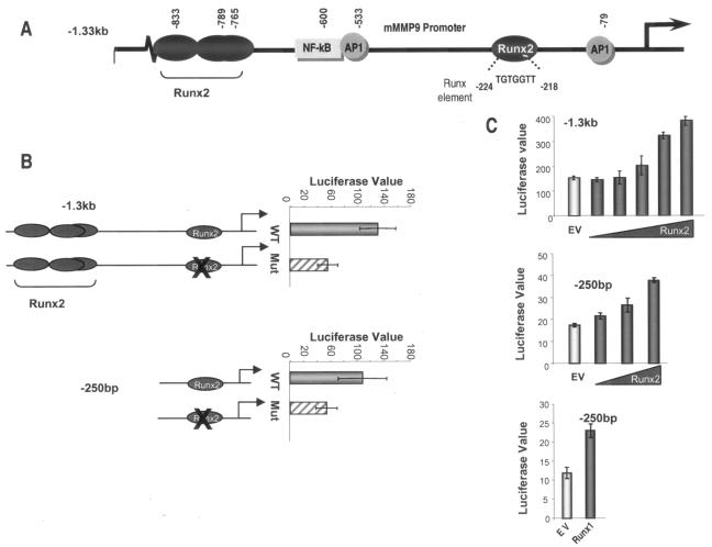
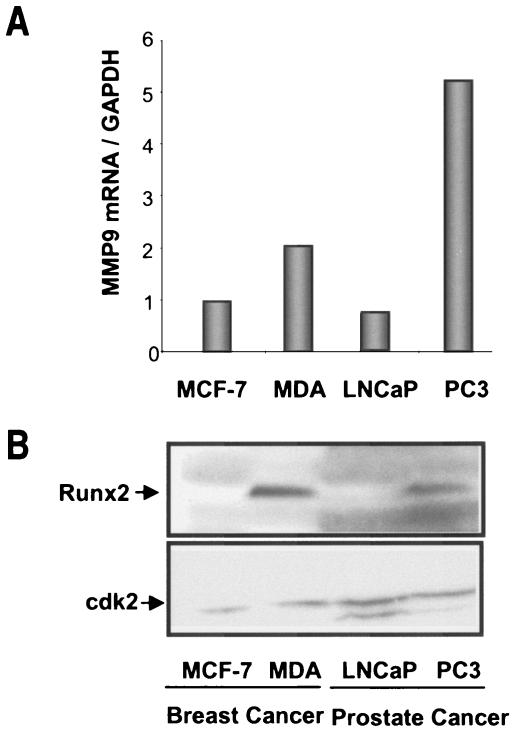
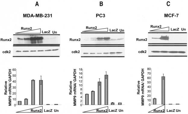
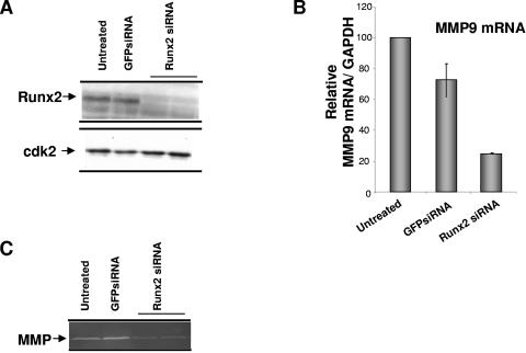
The Runx2 (Cbfa1/AML3) transcription factor and matrix metalloproteinase 9 (MMP9) are key regulators of growth plate maturation and bone formation. The genes for both proteins are characteristic markers of breast and prostate cancer cells that metastasize to bone. Here we experimentally addressed the compelling question of whether Runx2 and MMP are functionally linked. By cDNA expression array analysis, we identified MMP9 as a novel downstream target of Runx2. Like that of MMP13, MMP9 expression is nearly depleted in Runx2 mutant mice. Chromatin immunoprecipitation and electrophoretic mobility shift assays revealed the recruitment of Runx2 to the MMP9 promoter. We show by mutational analysis that the Runx2 site mediates transactivation of the MMP9 promoter in osteoblasts (MC3T3-E1) and nonosseous (HeLa) cells. The overexpression of Runx2 by adenovirus delivery in nonmetastatic (MCF-7) and metastatic breast (MDA-MB-231) and prostate (PC3) cancer cell lines significantly increases the endogenous levels of MMP9. The knockdown of Runx2 by RNA interference decreases MMP9 expression, as well as that of other Runx2 target genes, including the genes for MMP13 and vascular endothelial growth factor. Importantly, we have demonstrated using a cell invasion assay that Runx2-regulated MMP9 levels are functionally related to the invasion properties of cancer cells. These results are consistent with Runx2 control of multiple genes that contribute to the metastatic properties of cancer cells and their activity in the bone microenvironment.


Figures

FIG. 1.
FIG. 1.
MMP9 (also called gelatinase B) is a novel downstream target of Runx2. (A) Schematic illustration of domain organization of Runx2. QA, polyglutamate and -alanine stretch; RHD, runt homology domain; NLS, nuclear localization signal; NMTS, nuclear matrix targeting signal. (B) Total RNA (5 μg) from wild-type (WT) and Runx2ΔC/ΔC mutant mice (at 17.5 dpc) was hybridized to an osteogenesis-related cDNA array (left panel). The signals of MMP13 and -9 on blots are indicated in circles (middle panel). The MMP9 mRNA levels (normalized to glyceraldehyde-3-phosphate dehydrogenase [GAPDH]) in WT and Runx2ΔC/ΔC mutant mice were detected by RT-QPCR analysis (right panel). Protein extracts from the long bones of WT and Runx2ΔC/ΔC littermates were resolved by 8% SDS-PAGE with 2% gelatin and processed for zymography (C) and Western blotting (D) as described in Materials and Methods. (E) Immunolocalization of MMP9 in wild-type and Runx2ΔC/ΔC mutant embryos (at 17.5 dpc). Embryos were fixed in paraformaldehyde and embedded in paraffin, and the heads were serially sectioned at 8 μm. The left panels show MMP9 staining of the growth plate (GP) area of exoccipital bone. The ossification front (OF) is further magnified in the right panels showing the MMP9 signal. (The image was generated by overlaying the MMP9 image with an image that reflects sample autofluorescence. The MMP9 signal is shown in green, the background is shown in red, and autofluorescence is shown in yellow). Quantitation of the MMP9 signal in WT and Runx2 mutant tissue sections is shown by line scanning (the position is indicated in the micrograph) using Metamorph software, which measures the intensity of the fluorescent signal.
FIG. 2.
FIG. 2.
Overexpression of Runx2 results in transcriptional activation of MMP9 promoter. (A) Promoter region (−1.3 kb) of mouse MMP9 showing Runx binding elements at positions −224 (TGTGGTT), −765 (TGAGGTC), −789 (ACCCCAG), and −833 (ACCCCAA) and NF-κB and two AP1 binding sites. (B) Basal activities of wild-type (WT) and Runx binding mutant (Mut) full-length (−1.3 kb, upper panel) and proximal (−250 bp, lower panel) MMP9 promoters in mouse osteoblastic MC3T3-E1 cells. Cells were transfected with either the WT or mutant MMP9 promoter (200 ng/well in six-well plates) using the Fugene 6 reagent. (C) Effect of Runx2 expression on −1.3-kb MMP9 promoter (upper panel) and of Runx1 or Runx2 on −250-bp MMP9 promoter (middle and lower panels) in HeLa cells. Samples were analyzed for luciferase activity after 36 h of transient transfection with increasing amounts of Runx2 (200 ng to 1.6 μg) or Runx1 (1.6 μg) expression plasmid. EV, empty vector control for Runx2 expression construct. The promoter activity (luciferase value × 1,000) was normalized by cotransfection with Renilla luciferase.
FIG. 3.
FIG. 3.
Runx2 occupancy of MMP9 promoter. (A) Nuclear extracts were prepared from ROS 17/2.8 and primary rat osteoblasts (ROB) (day 12 [matrix maturation stage] and day 20 [mineralization]). An oligonucleotide containing the Runx binding element from the mouse MMP9 promoter (−212 bp to −246 bp) was incubated with 4 μg (lanes 3, 5, and 8) or 8 μg (lanes 6 and 9) of nuclear extract. The open arrow represents the specific DNA-protein complex, and the filled arrow shows a supershift with 1 μl of Runx2 polyclonal antibody (lanes 4, 7, and 10). (B) Competition assay performed with increasing amounts (25-, 50-, and 100-fold molar excess) of either cold wild-type (WT) or Runx mutant (mt) oligonucleotide showing the specificity of the Runx2-DNA complex. (C) In vivo occupancy of Runx2 protein at the proximal MMP9 promoter, as shown by ChIP of nuclei from day 12 ROB cells. The primers used to amplify the key regulatory elements present in the proximal MMP9 promoter fragment in the ChIP assays are indicated. The arrows indicate the positions of the forward (−380) and reverse (−130) primers. The agarose gel presented is representative of three experiments and shows selective amplification of the MMP9 proximal promoter in the input and Runx2 immunoprecipitate lanes, while IgG did not show any product.
FIG. 4.
FIG. 4.
Basal expression of MMP9 and Runx2 in nonmetastatic and bone metastatic cancer cells. (A) Total RNA from bone metastatic breast cancer cells (MDA-MB-231) and prostate cells (PC3), as well as from nonmetastatic breast (MCF-7) and prostate (LNCaP) tumor cells, was utilized to examine endogenous MMP9 mRNA levels by RT-QPCR analysis. Data were normalized to the GAPDH signal. (B) Whole-cell lysates from cancer cells were utilized to detect Runx2 protein with a monoclonal antibody by Western blotting. cdk2 protein levels are shown as an internal loading control.
FIG. 5.
FIG. 5.
Overexpression of Runx2 results in transcriptional activation of endogenous MMP9. Bone metastatic (MDA-MB-231 and PC3) and nonmetastatic (MCF-7) cancer cells were transduced with a LacZ- or Runx2-expressing adenovirus at increasing MOIs (for MDA-MB-231 and PC3 cells, 5, 10, 20, and 40 MOI; for MCF-7 cells, 10 and 40 MOI) for 48 h. Whole-cell lysates were utilized to detect the Runx2 protein with a mouse monoclonal antibody, and protein levels of cdk2 were measured as an internal loading control. Total RNA isolated from cells infected with the Runx2- or LacZ-expressing adenovirus (control) or from untreated (Un) cells was utilized to examine the MMP9 mRNA, as detected by RT-QPCR analysis. The amount of MMP9 mRNA was normalized to that of GAPDH.
FIG. 6.
FIG. 6.
Runx2 knockdown results in decreased endogenous MMP9 levels in metastatic breast cancer cells. (A) MDA-MB-231 breast cancer cells were treated with siRNAs for Runx2 (25 nM) and for GFP as a control with Oligofectamine for 72 h. Whole-cell lysates were analyzed for Runx2 protein and cdk protein as an internal loading control by Western blotting. (B) mRNA levels of endogenous MMP9 were detected by real-time RT-PCR of total RNA isolated from cells treated with siRNA for Runx2 or GFP or from untreated cells. (C) MMP9 protein activity was detected using gelatin zymography.
FIG. 7.
FIG. 7.
Runx2 influences invasion of metastatic and nonmetastatic cancer cells. (A) Untreated breast cancer (MDA-MB-231) cells (control) or cells treated with either Runx2 or GFP siRNA (25 nM) for 48 h (2 × 10⁵ cells) were plated on Matrigel or control inserts for an invasion assay. Cells were allowed to migrate for 20 h at 37°C. Cells that migrated were counted in 10 random fields, and the data are presented as percentages of invasion relative to control inserts. (B) MCF cells (4 × 10⁵ cells) infected with Runx2 or LacZ (control) adenovirus were plated on a Matrigel-coated filter (12-μm pore size) and allowed to migrate for 24 h at 37°C.
FIG. 8.
FIG. 8.
Runx2 knockdown decreases mRNA levels of metastatic markers (MMP2, MMP13, and VEGF) in MDA-MB-231 breast cancer cells. (A) Real-time RT-PCR analysis of total cellular RNA (5 μg) prepared from cells treated with Runx2 or GFP siRNA for 48 h. GAPDH mRNA was used as a control for normalization. (B) Transcriptional regulation of metastatic markers and Runx2 target genes known to be expressed in cancer cells. Breast cancer cells from primary tumors express metastatic markers (VEGF and MMPs) that are directly regulated by Runx2. It is not clear when during the metastatic process the Runx2 protein is first detected. The presence of Runx2 in cancer cells from bone metastatic tumors further activates the expression of tumor-related target genes.

References

    1. Aalinkeel, R., M. P. Nair, G. Sufrin, S. D. Mahajan, K. C. Chadha, R. P. Chawda, and S. A. Schwartz. 2004. Gene expression of angiogenic factors correlates with metastatic potential of prostate cancer cells. Cancer Res. 64:5311-5321. - PubMed
    1. Afzal, F., J. Pratap, K. Ito, Y. Ito, J. L. Stein, A. J. van Wijnen, G. S. Stein, J. B. Lian, and A. Javed. 2005. Smad function and intranuclear targeting share a Runx2 motif required for osteogenic lineage induction and BMP2 responsive transcription. J. Cell. Physiol. 204:63-72. - PubMed
    1. Amorino, G. P., and R. L. Hoover. 1998. Interactions of monocytic cells with human endothelial cells stimulate monocytic metalloproteinase production. Am. J. Pathol. 152:199-207. - PMC - PubMed
    1. Banerjee, C., L. R. McCabe, J.-Y. Choi, S. W. Hiebert, J. L. Stein, G. S. Stein, and J. B. Lian. 1997. Runt homology domain proteins in osteoblast differentiation: AML-3/CBFA1 is a major component of a bone specific complex. J. Cell. Biochem. 66:1-8. - PubMed
    1. Barnes, G. L., K. E. Hebert, M. Kamal, A. Javed, T. A. Einhorn, J. B. Lian, G. S. Stein, and L. C. Gerstenfeld. 2004. Fidelity of Runx2 activity in breast cancer cells is required for the generation of metastases associated osteolytic disease. Cancer Res. 64:4506-4513. - PubMed

Publication types

MeSH terms