. 2005 Oct;25(19):8683-92.
doi: 10.1128/MCB.25.19.8683-8692.2005.

pH-Responsive, posttranslational regulation of the Trk1 potassium transporter by the type 1-related Ppz1 phosphatase

Affiliations

pH-Responsive, posttranslational regulation of the Trk1 potassium transporter by the type 1-related Ppz1 phosphatase

Lynne Yenush et al. Mol Cell Biol. 2005 Oct.

Abstract

Intracellular pH and K+ concentrations must be tightly controlled because they affect many cellular activities, including cell growth and death. The mechanisms of homeostasis of H+ and K+ are only partially understood. In the yeast Saccharomyces cerevisiae, proton efflux is mediated by the Pma1 H+-ATPase. As this pump is electrogenic, the activity of the Trk1 and -2 K+ uptake system is crucial for sustained Pma1p operation. The coordinated activities of these two systems determine cell volume, turgor, membrane potential, and pH. Genetic evidence indicates that Trk1p is activated by the Hal4 and -5 kinases and inhibited by the Ppz1 and -2 phosphatases, which, in turn, are inhibited by their regulatory subunit, Hal3p. We show that Trk1p, present in plasma membrane "rafts", physically interacts with Ppz1p, that Trk1p is phosphorylated in vivo, and that its level of phosphorylation increases in ppz1 and -2 mutants. Interestingly, both the interaction with and inhibition of Ppz1p by Hal3p are pH dependent. These results are consistent with a model in which the Ppz1-Hal3 interaction is a sensor of intracellular pH that modulates H+ and K+ homeostasis through the regulation of Trk1p activity.


Figures

FIG. 1.
FIG. 1.
Trk1p is localized to plasma membrane rafts. Rafts were purified from the WΔ3 [pCM262-TRK1] yeast strain (grown in the presence of 20 μg/ml doxycycline) as described in Materials and Methods. The presence of Pma1p and the HA-tagged version of Trk1p in the various fractions was analyzed by Western blotting. Identical results were observed in four different experiments. The numbers above the lanes indicate the fraction number starting from the top of the gradient (lowest to highest density).
FIG. 2.
FIG. 2.
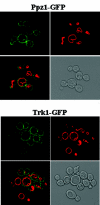
Localization of the Ppz1-GFP fusion protein. The strains LY 166 and LY 247 were analyzed by confocal microscopy, as described in Materials and Methods. The localization of the fusion proteins is shown in green. The FM4-64-stained vacuolar membranes are shown in red. Overlay and gray scale images are also shown.
FIG. 3.
FIG. 3.
Ppz1p partially associates with the lowest-density fraction in flotation gradients. Proteins were extracted from the W303-1A strain and processed as described in Materials and Methods. The Ponceau S staining of the membranes is shown in the top panel, as a control for protein loading. Fraction numbers are indicated above each lane (1 indicates the lowest-density and 8 indicates the highest-density fraction).
FIG. 4.
FIG. 4.
Redistribution of Ppz1p upon TRK1 overexpression. The indicated yeast strains (WΔ3, W303-1A, LY 152, and LY 161) were processed for flotation gradients as described in Materials and Methods. The top row shows the Ponceau S staining of the membranes as a control for protein loading. The amount of Ppz1p in each lane was estimated in digitally converted images using the MacBAS software. The LY 161 strain was grown in the presence (uninduced) or absence (induced) of doxycycline (20 μg/ml) to control TRK1 expression. Similar results were observed in two separate experiments. WT, wild type.
FIG. 5.
FIG. 5.
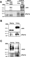
Ppz1p coimmunoprecipitates with Trk1p. Proteins were extracted from the indicated yeast strains, cross-linked, and processed for immunoprecipitation experiments as described in Materials and Methods. The proteins recovered by the affinity resin were analyzed by Western blotting using the indicated antibodies. In panel A, the first two lanes contain an aliquot of the crude extracts, and the last two lanes contain the immunoprecipitated material. In panels B and C, the lanes correspond to the immunoprecipitated material from the indicated strain.
FIG. 6.
FIG. 6.
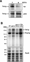
Phosphorylation of Trk1p in vitro and in vivo. (A) Proteins extracted from the yeast strain LY 236 were processed for immunoprecipitation experiments, followed by in vitro kinase assays, as described in Materials and Methods. On top are shown the results of the autoradiography, and the results of the Western blot used as a control of protein loading are below. Similar results were observed in three separate experiments. (B) The indicated strains were grown in low-phosphate media, and phosphorylated proteins were radioactively labeled in vivo and processed for raft purification, as described in Materials and Methods. Proteins present in the top fraction of the second gradient were separated by SDS-PAGE and analyzed by autoradiography (top). On the bottom is a Ponceau S-stained membrane showing equal amounts of protein present in each sample extracted just before labeling. Similar results were observed in three different experiments. WT, wild type.
FIG. 7.
FIG. 7.
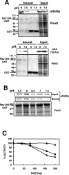
The in vitro interaction between Ppz1p and Hal3p is pH dependent. (A) Representative Western blot showing the amount of Hal3p (top) or Ypi1p (bottom) bound by the Ppz1ΔN-GST affinity resin at the pH indicated and the amount of Hal3p or Ypi1p in the starting extracts (input). The specificity of the binding interactions is demonstrated by the lack of binding to GST alone. Ponceau S-stained nitrocellulose membranes demonstrating equal protein loading are shown. The positions of GST and the Ppz1ΔN-GST fusion protein are indicated. Similar results were observed in two different experiments. (B) The same experiment as in panel A was performed at the indicated pHs. Shown are Western analysis with Hal3p-specific antibodies of the starting material (top) and the affinity-purified material (middle) and the Ponceau S-stained nitrocellulose membrane (bottom). (C) In vitro phosphatase assays were performed as described in Materials and Methods. The results are expressed as the percentage of phosphatase activity observed in the absence of Hal3p and represent the average of duplicate determinations (diamonds, pH 6.5; squares, pH 7.0; triangles, pH 7.5).
FIG. 8.
FIG. 8.
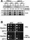
The pH dependence of the Ppz1-Hal3 interaction can be observed biochemically and phenotypically in vivo. (A) The amount of Hal3p recovered in the insoluble fraction was assayed as described in Materials and Methods. (Top) Representative Western analysis of the amount of Hal3p present in the indicated fractions. (Bottom) Ponceau S staining of the nitrocellulose filter as a control for equal protein loading in comparable fractions. (B) The indicated strains (LY 78, LY 79, and LY 83) were grown to saturation in rich media, serially diluted in water, and spotted on the indicated solid media. Growth was recorded after 48 to 72 h of incubation at 28°C.
FIG. 9.
FIG. 9.
Both Hal3p and the Ppz phosphatases are involved in the adaptation of yeast cells to acidic pH. The indicated strains were grown to saturation in rich media, serially diluted in water, and spotted on the indicated solid media. Growth was recorded after 48 to 72 h of incubation at 28°C. WT, wild type.

References

    1. Antz, C., T. Bauer, H. Kalbacher, R. Frank, M. Covarrubias, H. R. Kalbitzer, J. P. Ruppersberg, T. Baukrowitz, and B. Fakler. 1999. Control of K+ channel gating by protein phosphorylation: structural switches of the inactivation gate. Nat. Struct. Biol. 6:146-150. - PubMed
    1. Bagnat, M., A. Chang, and K. Simons. 2001. Plasma membrane proton ATPase Pma1p requires raft association for surface delivery in yeast. Mol. Biol. Cell 12:4129-4138. - PMC - PubMed
    1. Bagnat, M., S. Keranen, A. Shevchenko, A. Shevchenko, and K. Simons. 2000. Lipid rafts function in biosynthetic delivery of proteins to the cell surface in yeast. Proc. Natl. Acad. Sci. USA 97:3254-3259. - PMC - PubMed
    1. Benito, B., F. Portillo, and R. Lagunas. 1992. In vivo activation of the yeast plasma membrane ATPase during nitrogen starvation. Identification of the regulatory domain that controls activation. FEBS Lett. 300:271-274. - PubMed
    1. Bianchini, L., G. L'Allemain, and J. Pouyssegur. 1997. The p42/p44 mitogen-activated protein kinase cascade is determinant in mediating activation of the Na+/H+ exchanger (NHE1 isoform) in response to growth factors. J. Biol. Chem. 272:271-279. - PubMed

Publication types

MeSH terms