Review
. 2006 Apr;13(4):539-50.
doi: 10.1038/sj.cdd.4401769.

HATs and HDACs in neurodegeneration: a tale of disconcerted acetylation homeostasis

Affiliations
Review

HATs and HDACs in neurodegeneration: a tale of disconcerted acetylation homeostasis

R N Saha et al. Cell Death Differ. 2006 Apr.

Abstract

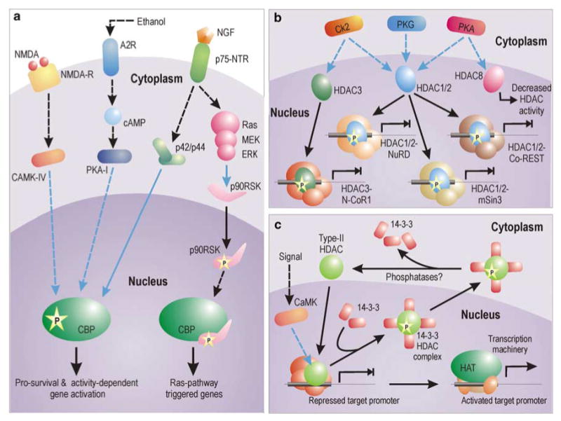
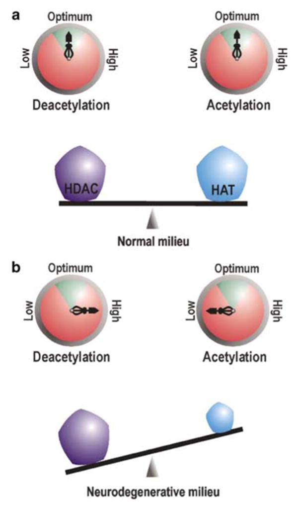
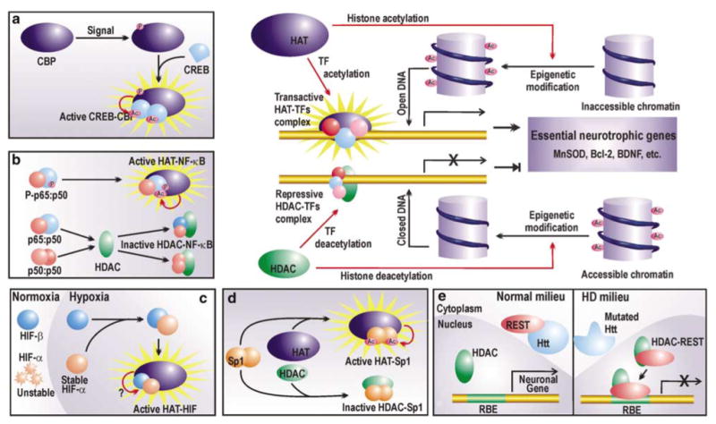
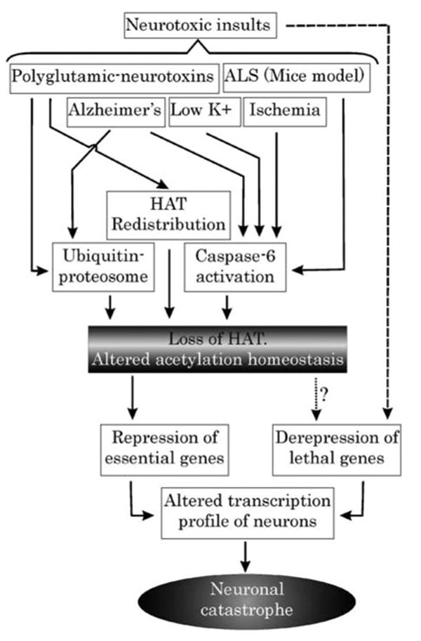
Gradual disclosure of the molecular basis of selective neuronal apoptosis during neurodegenerative diseases reveals active participation of acetylating and deacetylating agents during the process. Several studies have now successfully manipulated neuronal vulnerability by influencing the dose and enzymatic activity of histone acetyltransferases (HATs) and histone deacetylases (HDACs), enzymes regulating acetylation homeostasis within the nucleus, thus focusing on the importance of balanced acetylation status in neuronal vitality. It is now increasingly becoming clear that acetylation balance is greatly impaired during neurodegenerative conditions. Herein, we attempt to illuminate molecular means by which such impairment is manifested and how the compromised acetylation homeostasis is intimately coupled to neurodegeneration. Finally, we discuss the therapeutic potential of reinstating the HAT-HDAC balance to ameliorate neurodegenerative diseases.


Figures

Figure 1
Figure 1
Signaling mechanisms for the activation of HAT and HDAC. (a) Pathways for the activation of CBP HAT in neurons. Activity-dependent activation of CBP depends on CAMK-IV-dependent phosphorylation on Ser 301. CBP is also activated by elevation in cAMP level. Ethanol, for example, phosphorylates adenosine A2 receptor by blocking adenosine uptake and its buildup. Subsequently, cAMP level is increased by adenylyl cyclase. cAMP-mediated activation of CBP is dependent on type-I PKA-mediated phosphorylation. Growth factors (like NGF) can trigger CBP phosphorylation via p42/p44 MAPK, which has been shown to interact directly with CBP. NGF can also trigger Ras pathway to mediate phosphorylation of 90 kDa ribosomal S6 kinase (RSK), which binds to CBP and canalizes expression of genes dependent on Ras pathway. However, this interaction inhibits expression of genes with CRE elements in PC12 cells. (b and c) Different modus operandi of class-I and class-II HDACs. HDAC1, HDAC2 and HDAC3 are phosphorylated by a distinct set of kinases. Once phosphorylated, they assemble into greater repressive complexes showing greater enzymatic activity and inhibit gene expression. In contrast, phosphorylation of HDAC8 decreases its enzymatic activity. Among all the represented HDAC complexes, the HDAC1/2-Co-REST complex is neuron specific. On the other hand, as shown in myoblasts, several type-II HDACs, once phosphorylated, are exported from the nucleus in complex with 14-3-3 proteins. The binding of HDACs with 14-3-3 proteins results in their release from the repression complex, allowing expression of the gene. Whether similar mechanisms are applicable to neurons is yet to be verified. Blue connectors represent kinase activity
Figure 2
Figure 2
Neuronal acetylation homeostasis. Pentagons on the balance beam represent the protein level (dose) of HATs and HDACs, while compasses represent their activity. Enzymatic activity within the green arc of the compass is physiologically optimum. (a) In neurons under normal conditions, the dose and activity of HATs and HDACs are poised in a fine balance where they counteract each other to ensure physiological homeostasis. (b) During neurodegeneration, critical loss of HAT protein level ensue a rebated HAT dose and activity. This reclines the acetylation balance towards excessive deacetylation of target moieties
Figure 3
Figure 3
Regulation of chromatin and prosurvival TFs by HATs and HDACs. HATs acetylate lysine residues on nucleosomal histones to render the chromatin accessible to the transcription machinery, which then upregulates the expression of an array of neurotrophic factors. HDACs reverse the process by histone deacetylation. Furthermore, HATs and HDACs also regulate the activity of certain prosurvival TFs (ad). (a) CREB activity depends on its acetylation status. Phosphorylated CBP can acetylate CREB, which then undergoes conformational modification and forms a transactive dimer in association with the HAT. (b) The time and strength of NF-κB p65 : p50 is mediated by acetylation of p65 by HATs. However, if unphosphorylated, NF-κB p65 : p50 can interact with HDACs to form nontransactive complexes. Furthermore, p50 : p50 can form repressive complex with HDACs. (c) Hypoxic responses of HIF require steric interaction of HATs with HIF heterodimers. However, the probable enzymatic HAT activity is not yet reported. (d) Sp1 may conditionally interact with HATs or HDACs. Transactivation by Sp1 is accrued by its interaction with HATs and subsequent acetylation. (e) Nonepigenetic repression by HDAC. During HD, HDAC represses the expression of vital neuronal genes by forming a repressor complex with REST, which otherwise in normal conditions is arrested in the cytoplasm by wild huntingtin protein (Htt). Refer to the text for abbreviations
Figure 4
Figure 4
Proposed model depicting central role of acetylation homeostasis during neurodegeneration: Various models and diseases of neurodegeneration are marked by critical loss of HAT. This loss, leading to acetylation imbalance, may be manifested by various means, which are employed singularly or in combination during a particular disease. Loss of acetylation homeostasis forms the last common port where several neurotoxic insults converge to mediate a self-demolishing neuronal response. Such responses include repression of prosurvival genes on the one hand and derepression of lethal genes on the other

References

    1. Taunton J, Hassig CA, Schreiber SL. A mammalian histone deacetylase related to the yeast transcriptional regulator Rpd3p. Science. 1996;272:408–411. - PubMed
    1. Timmermann S, Lehrmann H, Polesskaya A, Harel-Bellan A. Histone acetylation and disease. Cell Mol Life Sci. 2001;58:728–736. - PMC - PubMed
    1. Marmorstein R, Roth SY. Histone acetyltransferases: function, structure, and catalysis. Curr Opin Genet Dev. 2001;11:155–161. - PubMed
    1. Thiagalingam S, Cheng KH, Lee HJ, Mineva N, Thiagalingam A, Ponte JF. Histone deacetylases: unique players in shaping the epigenetic histone code. Ann N Y Acad Sci. 2003;983:84–100. - PubMed
    1. LaMorte VJ, Dyck JA, Ochs RL, Evans RM. Localization of nascent RNA and CREB binding protein with the PML-containing nuclear body. Proc Natl Acad Sci USA. 1998;95:4991–4996. - PMC - PubMed

Publication types