. 2005 Oct;115(10):2822-31.
doi: 10.1172/JCI24156. Epub 2005 Sep 15.

An interstitial deletion-insertion involving chromosomes 2p25.3 and Xq27.1, near SOX3, causes X-linked recessive hypoparathyroidism

Affiliations

An interstitial deletion-insertion involving chromosomes 2p25.3 and Xq27.1, near SOX3, causes X-linked recessive hypoparathyroidism

Michael R Bowl et al. J Clin Invest. 2005 Oct.

Abstract

X-linked recessive hypoparathyroidism, due to parathyroid agenesis, has been mapped to a 906-kb region on Xq27 that contains 3 genes (ATP11C, U7snRNA, and SOX3), and analyses have not revealed mutations. We therefore characterized this region by combined analysis of single nucleotide polymorphisms and sequence-tagged sites. This identified a 23- to 25-kb deletion, which did not contain genes. However, DNA fiber-FISH and pulsed-field gel electrophoresis revealed an approximately 340-kb insertion that replaced the deleted fragment. Use of flow-sorted X chromosome-specific libraries and DNA sequence analyses revealed that the telomeric and centromeric breakpoints on X were, respectively, approximately 67 kb downstream of SOX3 and within a repetitive sequence. Use of a monochromosomal somatic cell hybrid panel and metaphase-FISH mapping demonstrated that the insertion originated from 2p25 and contained a segment of the SNTG2 gene that lacked an open reading frame. However, the deletion-insertion [del(X)(q27.1) inv ins (X;2)(q27.1;p25.3)], which represents a novel abnormality causing hypoparathyroidism, could result in a position effect on SOX3 expression. Indeed, SOX3 expression was demonstrated, by in situ hybridization, in the developing parathyroid tissue of mouse embryos between 10.5 and 15.5 days post coitum. Thus, our results indicate a likely new role for SOX3 in the embryonic development of the parathyroid glands.


Figures

Figure 1
Figure 1
Identification of a less than 28-kb deletion on chromosome Xq27.1 in X-linked recessive HPT patients. The disease locus is located between the SNP rs5907612 and the microsatellite polymorphism DXS984 (9), which are shown on the expanded view alongside the schematically represented X chromosome with Giemsa bands. This 906-kb critical interval contains 3 genes — ATP11C, U7snRNA, and SOX3 — and abnormalities involving the coding regions of these genes in X-linked recessive HPT patients has been previously excluded (9). To further reduce the critical interval, SNPs from the telomeric boundary were initially sought, and analysis of the contig comprising BACs RP11-359I11, RP11-51C14, and RP4-595A18 revealed 2 SNPs, rs732125 and rs461191, that were respectively located approximately19 kb and 8 kb centromeric to U7snRNA. SNP rs461191 proved uninformative in the X-linked recessive HPT kindreds. However, analysis with SNP rs732125 revealed a likely deletion, as PCR products were not obtained from the 6 affected males (filled square) but were obtained from the 16 unaffected males (open square), 11 carrier females (circle with a dot), and 28 unaffected females (data not shown). This deletion was further delineated using 5 STSs (a–e) that were designed at intervals along the sequence of BAC RP11-51C14. PCR products were obtained using STSs a and e but not STSs b, c, and d, thereby indicating that the deletion extended from STS a to STS e, an interval of less than 28 kb.
Figure 2
Figure 2
Identification of a large insertion on chromosome Xq27.1 in X-linked recessive HPT patients. In order to confirm the less than 28-kb deletion involving STSs b, c, and d and SNP rs732125 (Figure 1) in X-linked recessive HPT patients, PFGE restriction mapping studies (20) were undertaken. (A) The less than 28-kb deletion predicted the occurrence of an approximately 250-kb SfiI fragment in X-linked recessive HPT patients, as compared with an approximately 275-kb SfiI fragment in normal individuals, when a [32P]-labeled SOX3-derived probe for hybridization was used. (B) PFGE confirmed the presence of an approximately 275-kb SfiI fragment in normal males (open square) but revealed an unexpected larger SfiI fragment of approximately 325 kb in males affected with X-linked recessive HPT (filled square). Carrier females (circle with a dot) had both the approximately 275-kb and 325-kb SfiI fragments. The results from 3 members of family W/81 (5, 8), which consists of 48 members, are shown, and the individual’s identification code regarding generation number and position in the pedigree is as previously reported (8) to facilitate comparison between the studies. (C) These findings are consistent with the occurrence of a large, at least 75-kb [i.e., (325–275) + 28 kb] insertion at the site of the less than 28-kb deletion (del) in individuals affected with X-linked recessive HPT.
Figure 3
Figure 3
Characterization of the deletion-insertion in X-linked recessive HPT. (A) The DNA sequences of the centromeric and telomeric breakpoints (Point-cen and Point-tel, respectively) were determined using genomic libraries generated from EBV-lymphoblastoid DNA and flow-sorted X chromosomes of an affected male (22). The DNA sequence from the centromeric boundary consisted of a dinucleotide repeat, and the DNA sequence from the telomeric boundary was therefore used to design ISPs (ISP1, ISP2, and ISP3) and XSPs (XSPF and XSPR) for the further characterization of the insertion. The sizes of the PCR products obtained with each primer pair are indicated. The locations of STSs a–e (Figure 1) are shown. (B) Use of primers ISP1 and ISP2 and DNA from a monochromosomal somatic cell hybrid panel revealed that the insert sequence originated from chromosome 2. 1+X, DNA from cell with chromosomes 1 and X; Hs, human DNA; Mm, mouse DNA; Ha, hamster DNA; –, no DNA. (C) Use of primers ISP3 and XSPR, which flank the telomeric breakpoint, identified the deletion-insertion in affected males (filled squares) and carrier females (circles with a dot). Use of primers XSPF and XSPR identified the normal X chromosome allele in unaffected males (open squares), unaffected females (open circles), and carrier females. The results from 8 members of family W/81 are shown, and the identification code used for individuals is as described in Figure 2. Individual 7a, who is an unaffected sister, has not been reported in previous studies (8). The combined results from all the members of families W/81(8) and P/60 (8) yielded a LOD score of 8.1 at 0% recombination.
Figure 4
Figure 4
FISH analyses confirm the deletion-insertion involving Xq27.1 and 2p25 in X-linked recessive HPT. (A) Metaphase-FISH analysis, using 2p25 fosmid AG63A8 (green signal) and Xq27 BAC RP11-51C14 (red signal). Chromosomes counterstained with DAPI (blue), and arrows indicate chromosomes 2 and X, as well as and abnormal X containing insert from 2p25 (X+2). Insets show enlarged views of X chromosome. Juxtaposed signals of fosmid and BAC, at Xq27, due to the deletion-insertion, yielded the yellow color. (B) Use of 3-color DNA fiber–FISH analyses revealed a large insertion that was derived from 2p25, at Xq27 in affected males. DNA fibers were hybridized with 2p25 YAC 972C12 (size, 750 kb), and the Xq27 BACs (Figure 1) RP11-51C14 (size, 149 kb) and RP11-359I11 (size, 43 kb). YAC 972C12 was labeled with fluorescein, BAC RP11-51C14 with Alexa-594, and BAC RP11-359I11 with Cy5, to yield green, red, and far red (pseudocolored in blue) signals, respectively. The numbers of observed DNA fibers yielding signals are indicated. (C) Contig of BACs (solid bars) and fosmid (broken bar) for YAC 972C12. (D) BACs were labeled with Texas red and separately hybridized to metaphase chromosome spreads. Two red signals corresponding to 2p25 hybridizations (white arrows) were obtained for each BAC (data shown for BACs RP11-97B21, CTD-2029L3, and RP13-542C4), and a third signal corresponding to Xq27 hybridizations (yellow arrows) was also detected for BACs CTD-2029L3, RP11-1268F2 (data not shown), and RP13-542C4. These results indicate that the insertion at Xq27.1 is contained between CTD-2029L3 and RP13-542C4 and is approximately 305–340 kb in size. The identification code used for individuals is as described in Figure 2.
Figure 5
Figure 5
Molecular mechanism underlying the deletion-insertion abnormality involving chromosomes Xq27 and 2p25 in X-linked recessive HPT. The relationships of the deletion-insertion to that of BACs (Figures 1 and 4) from chromosomes 2p25 (CTD-2029L3, RP11-1268F2, XXfos-83269E11, RP13-932E12, RP11-1294H20, and RP13-542C4) and Xq27 (RP11-51C14) are shown. DNA sequence database analyses together with a characterization of the DNA sequences of the breakpoints (Figures 2 and 3) in X-linked recessive HPT patients established that the deletion within Xq27.1 extends approximately 23–25 kb and that the insertion from chromosome 2p25.3, which inverted upon insertion, extends approximately 305–340 kb and includes exons 2–16 of the SNTG2 gene. The location of this deletion-insertion is approximately 67 kb downstream of the gene encoding SOX3, which belongs to a family of transcription factors that are involved in vertebrate embryonic development (13). For example, abnormalities of SOX3 expression are associated with developmental anomalies involving the CNS, craniofacial bones, and pituitary (–18, 41), while abnormalities of SRY expression are associated with disorders of sexual development (49). It is likely that the deletion-insertion causes a disruption of SOX3 regulatory elements and thereby exerts a position effect on SOX3 expression.
Figure 6
Figure 6
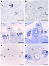
Expression of Sox3 and Casr in the developing parathyroids of mouse embryos. Transverse sections through the regions of the pharyngeal pouches and neck, from wild-type (normal) mouse embryos between 10.5 and 18.5 dpc, were hybridized with either Casr or Sox3 riboprobes. Casr, which is known to be expressed in the parathyroids and thyroids (6), was used as a control. (A) Casr expression was observed in the third pharyngeal pouch (Pp3) at 10.5 dpc. (B) On a serial section, Sox3 expression was observed in the posterior margins of the second (Pp2) and third pharyngeal pouch endoderm and also the foregut endoderm (Fg). (C) Sox3 and Casr (data not shown) expression were detected in the thyroid (Th) and in the thymic (Tm) and parathyroid (Pa) rudiments at 13.5 dpc. (D) Sox3 expression continued in the developing thyroid and parathyroids at 15.5 dpc. (E) However, Sox3 expression had almost ceased in the developing thyroid and parathyroids by 18.5 dpc. (F) In contrast, Casr was seen to be strongly expressed in the developing thyroid and parathyroids on a serial section at 18.5 dpc. Oe, esophagus; Tr, trachea. These results demonstrate that Sox3 is expressed, between 10.5 dpc and 15.5 dpc, in the pharyngeal pouches and developing parathyroids of the mouse embryo. Scale bars, 0.25 mm.

References

    1. Thakker RV. Genetics of endocrine and metabolic disorders: parathyroid. Rev. Endocr. Metab. Disord. 2004;5:37–51. - PubMed
    1. Baldini A. DiGeorge’s syndrome: a gene at last. Lancet. 2003;362:1342–1343. - PubMed
    1. Parkinson DB, Thakker RV. A donor splice site mutation in the parathyroid hormone gene is associated with autosomal recessive hypoparathyroidism. Nat. Genet. 1992;1:149–152. - PubMed
    1. Peden VH. True idiopathic hypoparathyroidism as a sex-linked recessive trait. Am. J. Hum. Genet. 1960;12:323–337. - PMC - PubMed
    1. Whyte MP, Weldon VV. Idiopathic hypoparathyroidism presenting with seizures during infancy: X-linked recessive inheritance in a large Missouri kindred. J. Pediatr. 1981;99:608–611. - PubMed

Publication types

MeSH terms